This is part of My Health, where the whole story is. This page has the scans of my spine and brain: the September 2026 MRIs, my April 2026 CBCT’s view of my upper neck sitting up, and what I make of my neck. A lumbar MRI of my lower back is booked for 27 October 2026, and its results will go here.
The September 2026 MRIs
On 21 September 2026 I had MRIs of my brain, cervical spine (neck) and thoracic spine (mid-back), each with and without contrast.
The figures below were annotated from the scan files. They aren’t a radiologist’s report; where I quote the report, I say so. Tap any figure to see it full size.
The short version: nothing is pressing on my spinal cord or nerves. According to these annotations, there’s no Chiari malformation, no enlarged ventricles and no low-lying or tethered cord. Two white-matter spots that looked like possible lesions turned out to be normal folds of brain. What did show up (a few small disc bulges and a small fatty lump in the brain) is small and common. And lying flat, my C1–C2 joint is about 5°, against about 18° sitting up in the CBCT, which tells me the rotation is functional rather than built into the bones.
Whole spine
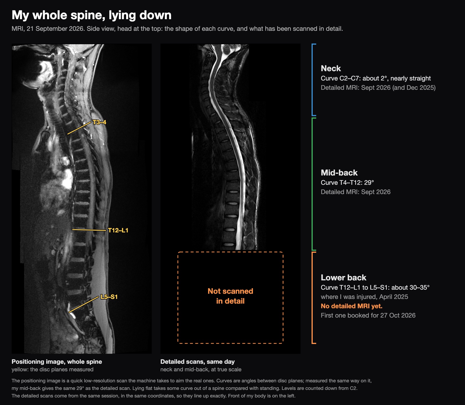
The scanner’s quick positioning image happens to cover my whole spine, top to bottom, so it’s the only look so far at my lower back. It’s low resolution and only a side view: it shows the shape of my spine, not the discs or nerves in any detail.
Lying flat, the shape is:
- Neck: about 2° from C2 to C7, so nearly straight (figure 8). That’s common on MRI, where you lie with your head in a coil.
- Mid-back: 29° from T4 to T12, inside the normal range of roughly 10–40°. Measured the same way on the positioning image, it also comes out at 29°, so I trust that image for the lower back too.
- Lower back: about 30–35° between the T12–L1 and L5–S1 discs. The top of my lower back, L1 and L2, stacks nearly straight, and most of the curve comes from the lowest few discs, which is where most people’s lower-back curve is. Standing, the usual range is roughly 40–60°. Lying flat takes some of that out, and measuring between discs rather than bone edges reads a few degrees lower again. So mine sits around the low end of normal: not obviously flat, and not overly arched.
At this resolution, nothing in my lower back looks collapsed or slipped out of line. That’s as far as this image goes. It can’t show a disc bulge or a pinched nerve, and as a side view it can’t show a sideways curve; the cross-sections found none from T1 to L1 (figure 4), and the lumbar MRI on 27 October 2026 is for the rest. And because I’m lying down, it says nothing about how I stand. The photos on How My Body Moves are better for that.
Mid-back
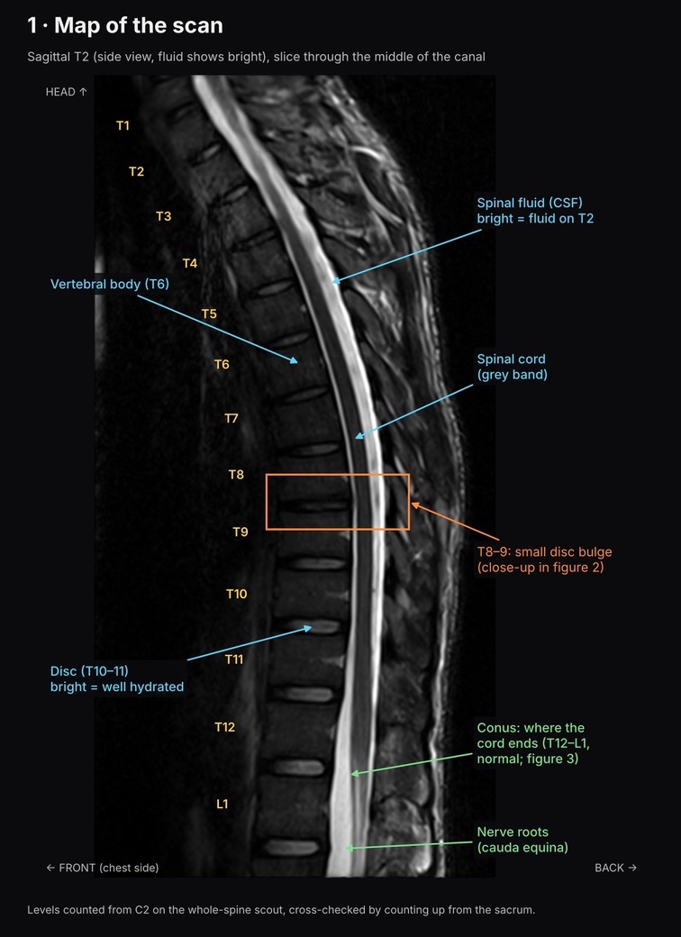
A side view of my mid-back, with fluid showing bright. The discs are well hydrated and the cord ends where it should. The one finding is a small bulge at T8–9.
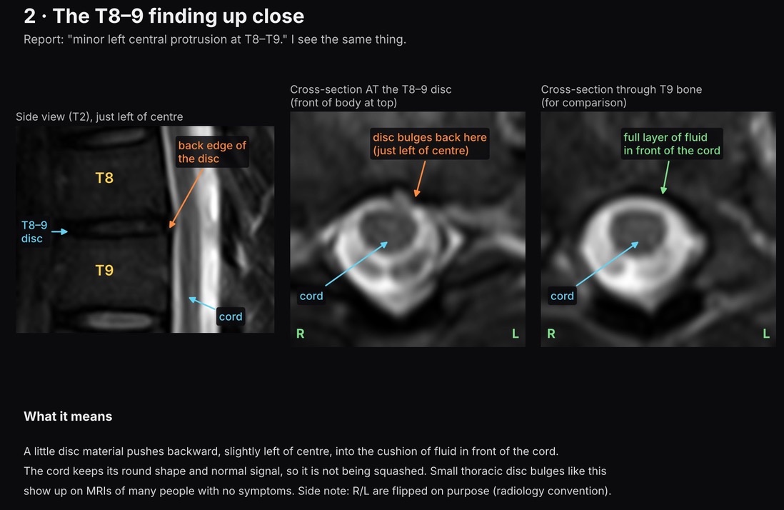
The radiology report calls it a “minor left central protrusion at T8–T9.” A little disc material pushes backward, just left of centre, into the cushion of fluid in front of the cord. The cord keeps its round shape and normal signal, so it isn’t being squashed.
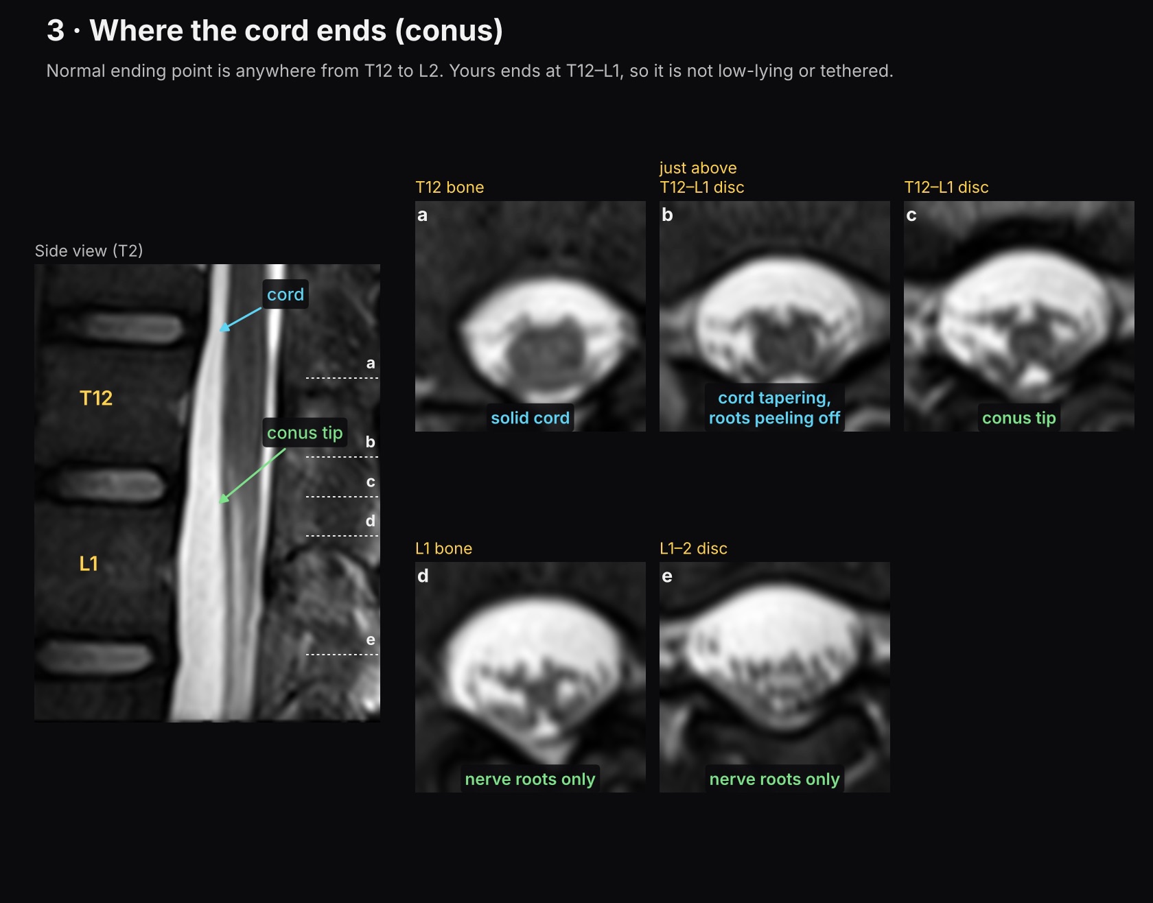
The cord normally ends anywhere from T12 to L2. Mine ends at T12–L1, so it isn’t low-lying or tethered.
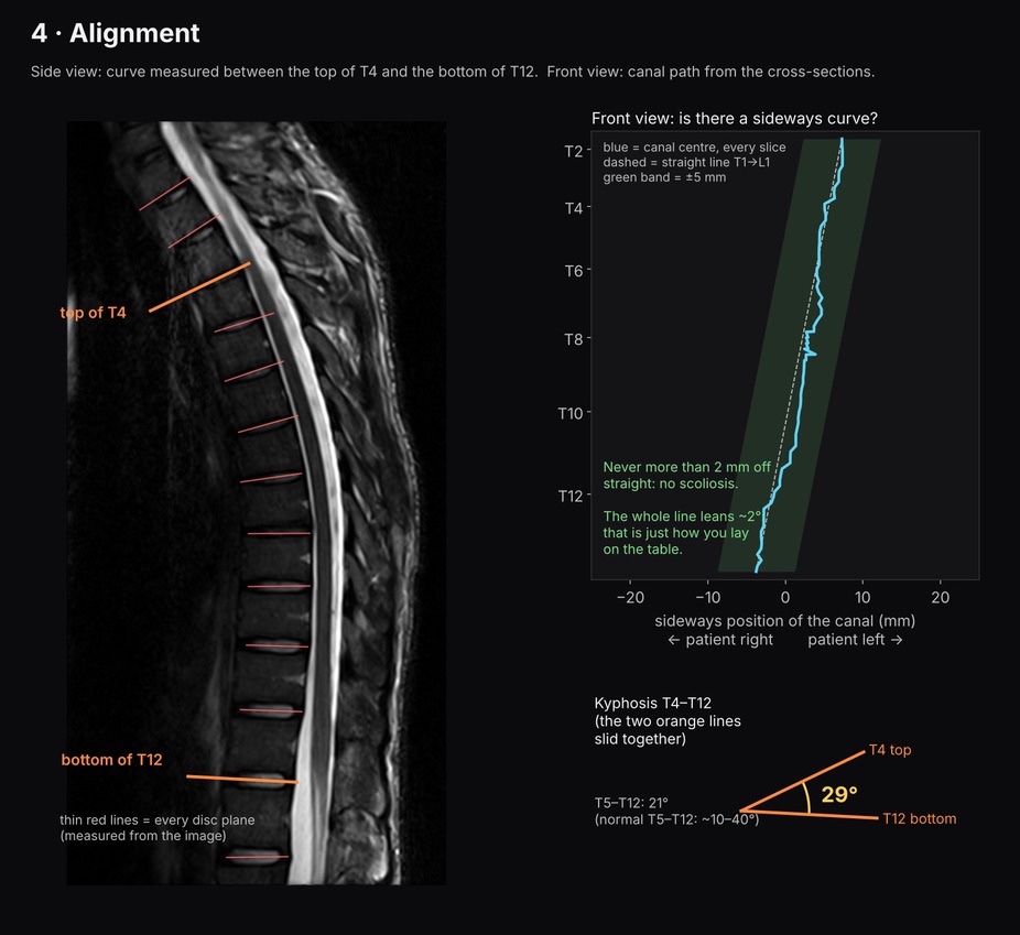
No sideways curve: the canal never strays more than 2 mm from a straight line, so there’s no scoliosis. The curve of my mid-back measures 29° from T4 to T12, and 21° from T5 to T12, where normal is roughly 10–40°.
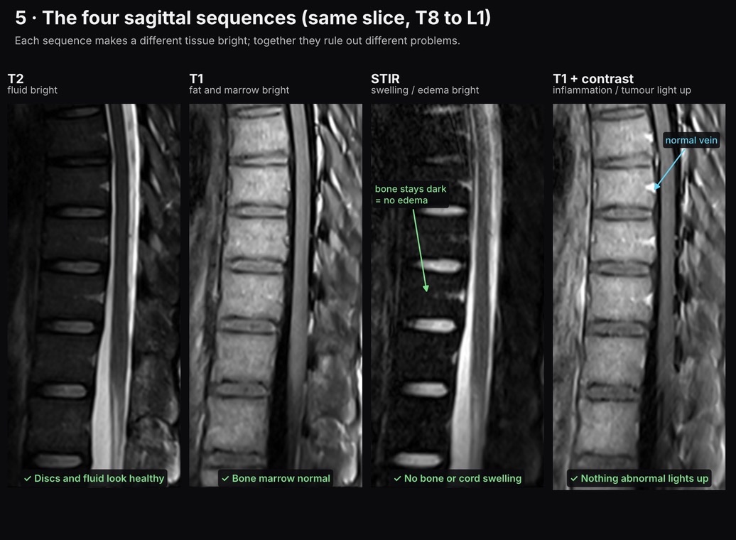
The same slice on four MRI sequences, each of which makes a different tissue bright. The discs and fluid look healthy, the bone marrow is normal, there’s no swelling in bone or cord, and nothing abnormal lights up with contrast.
Neck
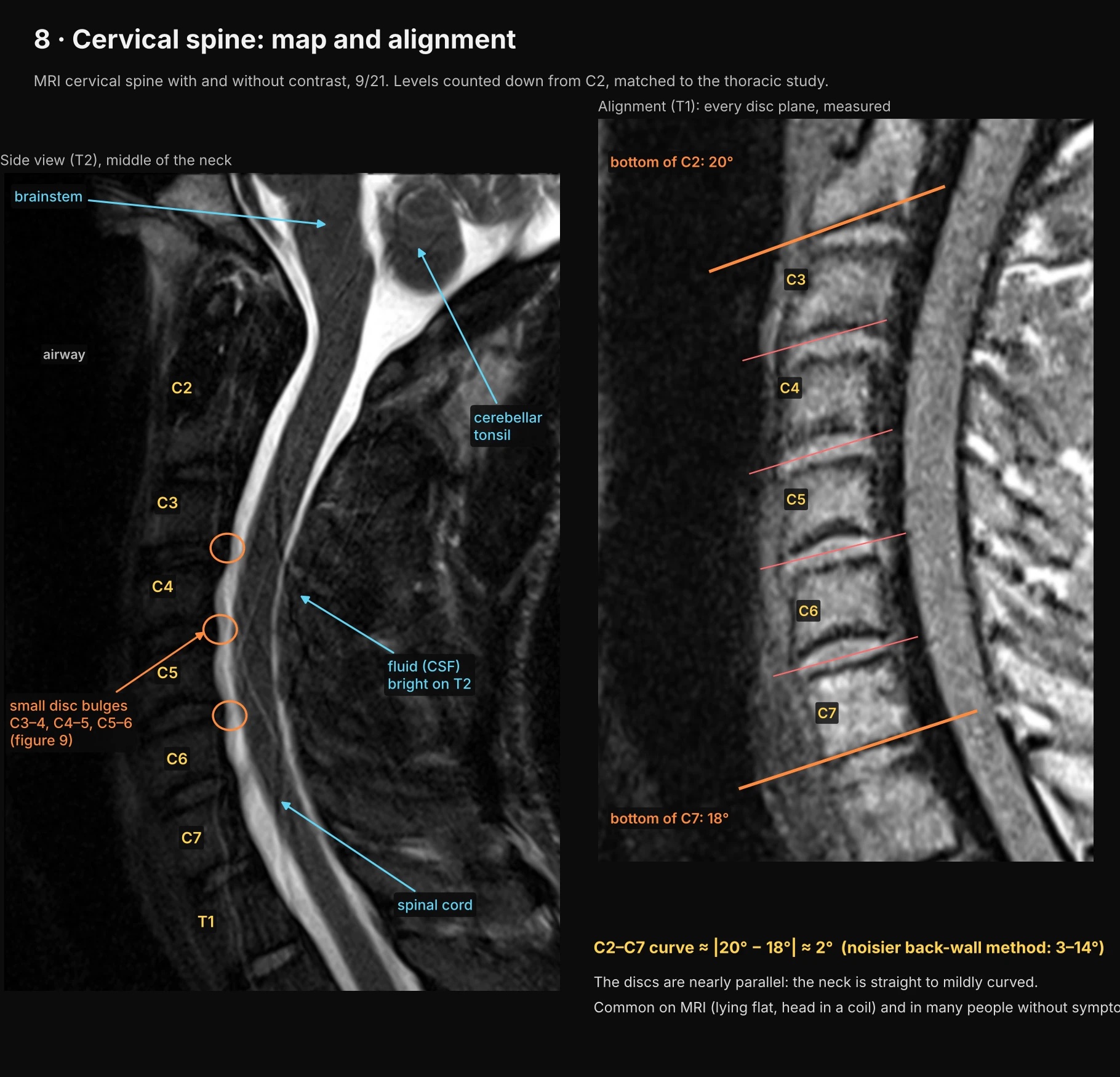
Small disc bulges at C3–4, C4–5 and C5–6. The curve of my neck from C2 to C7 measures only about 2°, so it’s straight to mildly curved. The annotations note that’s common on MRI, where you lie flat with your head in a coil, and in many people without symptoms.
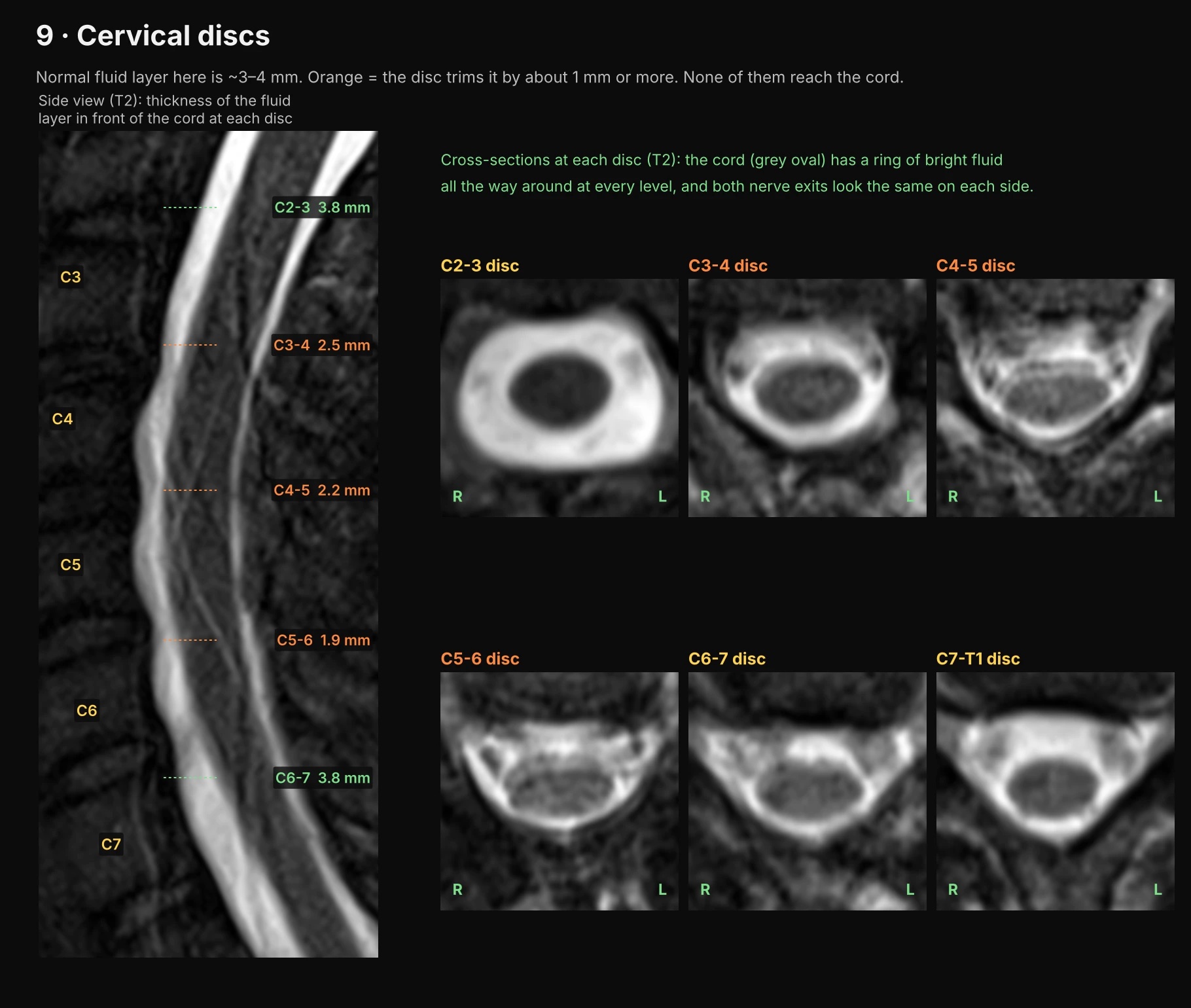
The layer of fluid in front of the cord is normally 3–4 mm thick. The three bulging discs trim it to 2.5, 2.2 and 1.9 mm, but none of them reaches the cord. The cord has a full ring of fluid at every level, and the nerve exits look the same on both sides.
Is C2–C4 rotated?
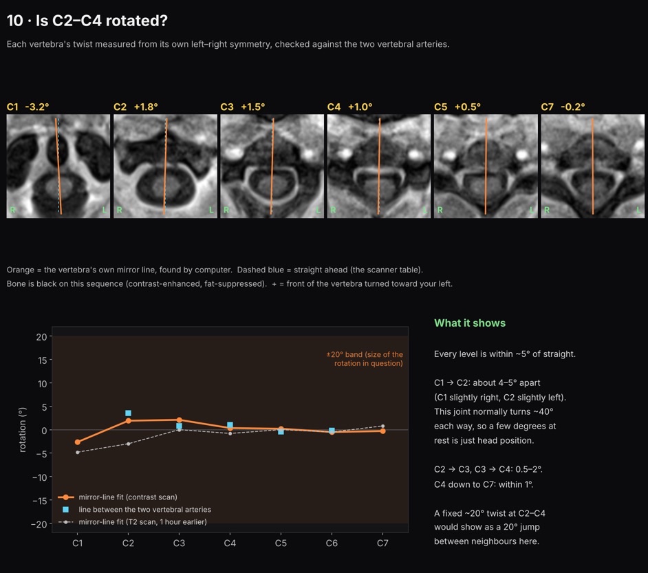
This is the figure that matters most to me. Each vertebra’s twist was measured from its own left-right symmetry and checked against the two vertebral arteries. Every level is within about 5° of straight. C1 and C2 are 4–5° apart, a joint that normally turns about 40° each way, so the annotations put that down to head position. A fixed 20° twist at C2–C4 would show up as a 20° jump between neighbours, and there isn’t one.
That fits with the CBCT rather than contradicting it. Sitting up after a long drive, the same C1–C2 joint measures about 18° (figure C); lying flat here, with my head held in a coil, it’s about 5°. So the rotation is functional rather than structural.
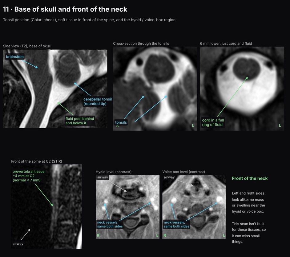
The base of my skull and the front of my neck. The cerebellar tonsils have rounded tips and sit where they should, so there’s no Chiari malformation. The tissue in front of the spine at C2 is about 4 mm thick, where normal is under 7. At the hyoid and voice box, the left and right sides look alike, with no mass or swelling. That’s the area where I feel the swallowing pain on the right, though this scan isn’t built for those tissues and can miss small things.
Upper neck, sitting up
This one is from my April 2026 CBCT, taken sitting up right after a long drive. The rest of that scan is on My Airway, Tongue and Jaw.
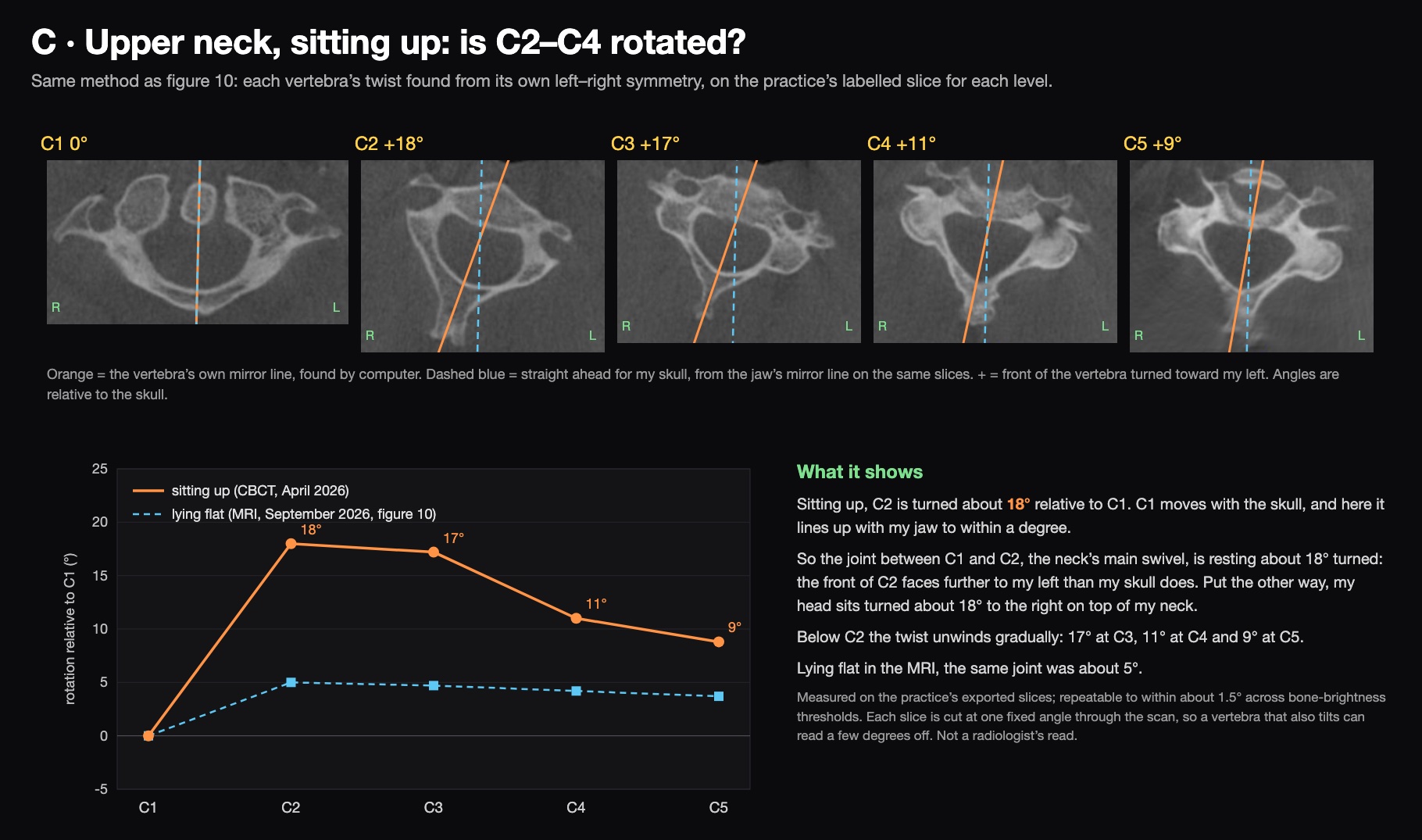
Each vertebra was measured the same way as in figure 10: find the line that best mirrors the bone onto itself, on the practice’s labelled slice for each level. C1 lines up with my jaw, so it’s turning with my skull. C2 is turned about 18° from it, with the front of C2 facing further to my left: seen from above, C2 is turned counterclockwise under my head. Put the other way, my head sat turned about 18° to the right on C2. A scan only shows how bones sit relative to each other, so those are the same fact. Below C2 the twist tapers: 17° at C3, 11° at C4, 9° at C5. So, measured against my skull, my whole neck is turned left, most at C2; put the other way, my head sits turned right on top of it. Lying flat in the MRI, the same C1–C2 joint was about 5°. On the front slices through the top of the neck, the peg of C2 also sits about 2 mm off-centre between the two sides of C1. That fits, but an offset that small is common, so it’s weak support on its own.
The angles repeat to within about 1.5° when the brightness a pixel needs to count as bone is changed. The bigger uncertainty is that these are 2D slices cut at one fixed angle through the scan, so a vertebra that also tilts can read a few degrees off.
Brain
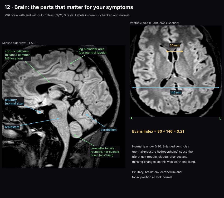
My ventricles are normal size: the Evans index, their width divided by the width of the skull, is 0.21, where normal is under 0.30. The pituitary, brainstem, cerebellum and tonsil position all look normal, and the corpus callosum, a common place for MS lesions, is clean.
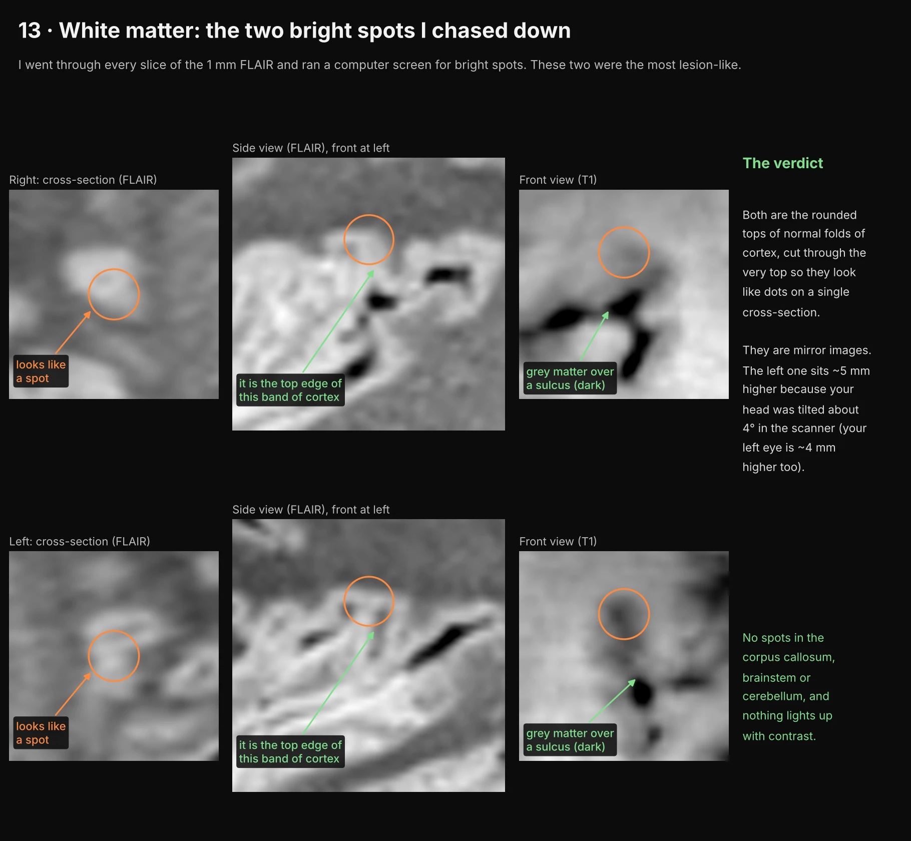
Two bright spots in the white matter looked like they could be lesions. Followed through the slices, both turn out to be the rounded tops of normal folds of cortex, cut so they look like dots on a single slice. There are no spots in the corpus callosum, brainstem or cerebellum, and nothing lights up with contrast.
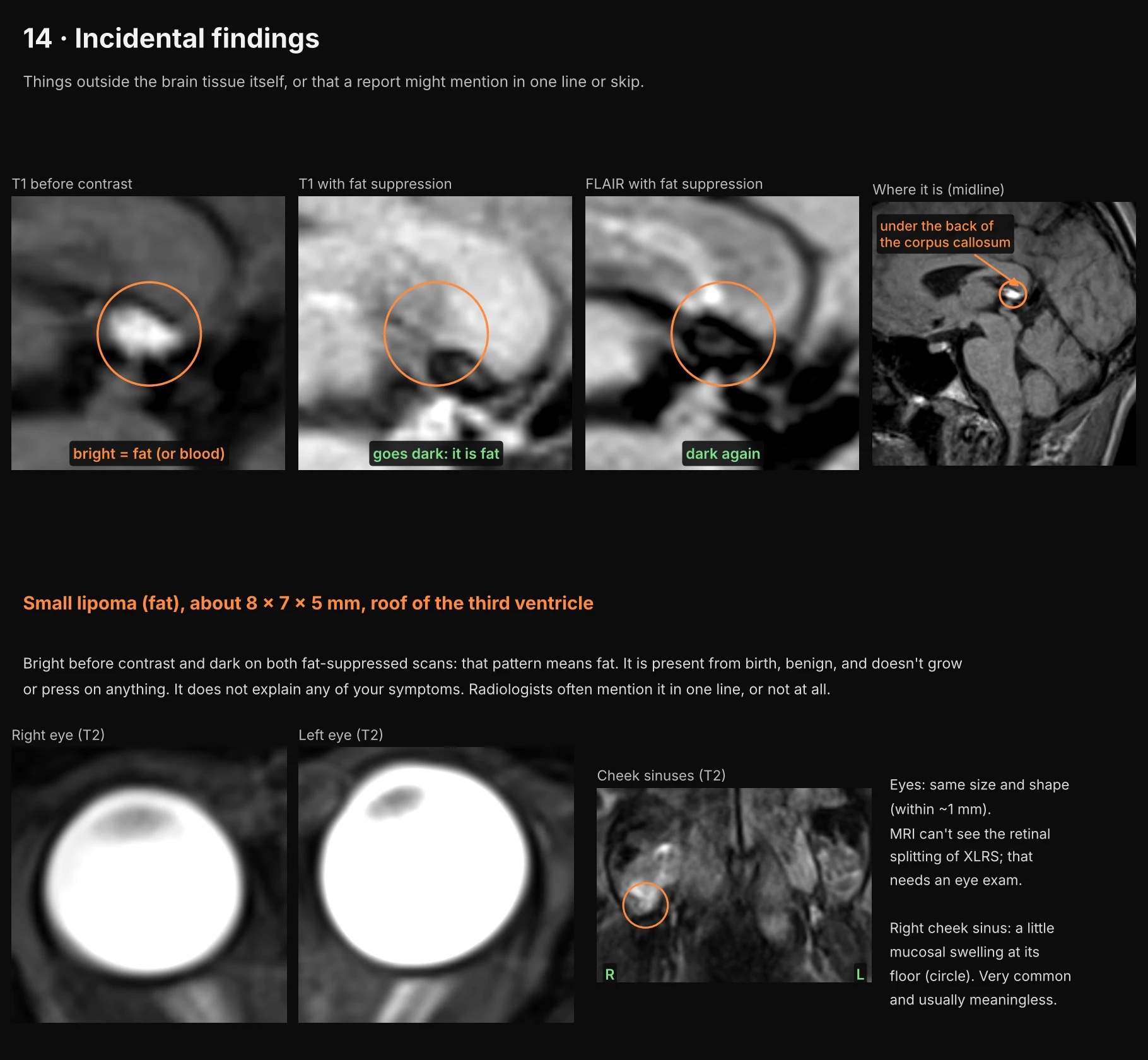
Two incidental findings. One is a small fatty lump (a lipoma, about 8 × 7 × 5 mm) on the roof of the third ventricle. It’s present from birth, benign, doesn’t grow or press on anything, and doesn’t explain any of my symptoms. The other is a little swelling in the lining of my right cheek sinus, which is very common. My eyes are the same size and shape.
My neck
I’d been told my upper neck is rotated about 20 degrees between the C2 and C4 vertebrae, and my April 2026 CBCT read noted visible rotation at C2.
The two scans I have were taken in different positions, and they disagree in a way that makes sense:
- Sitting up (the CBCT, right after the drive from Lebanon to Northwood, NH: upright, and straight after the kind of long sit that aggravates me): C2 sits about 18° turned relative to C1. See figure C.
- Lying flat (the MRI, with my head held in a coil): the same joint is about 5°, and no level is more than about 5° from straight. See figure 10.
I read the two together: the rotation is functional. It shows up in some states and not others, and it isn’t built into the bones. Both scans held my head in place, though (a chin rest for the CBCT, a head coil for the MRI), and they were measured by different methods. So this compares position, device and state together, not gravity alone, and the CBCT came straight after a long drive.
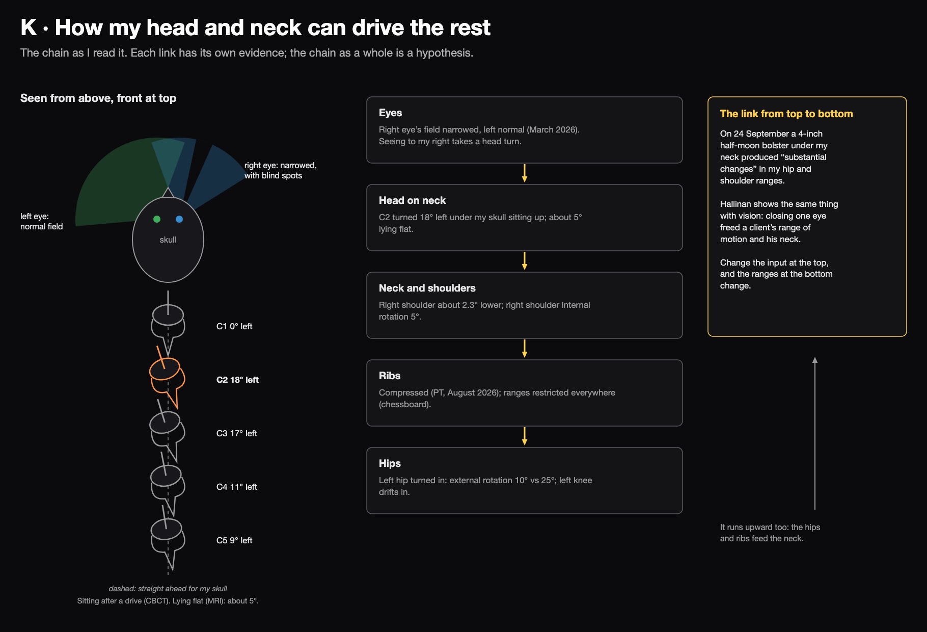
The chain from my eyes down to my hips is a hypothesis, but each link has its own evidence. The strongest piece is the bolster: changing something under my neck changed the ranges at my hips and shoulders.