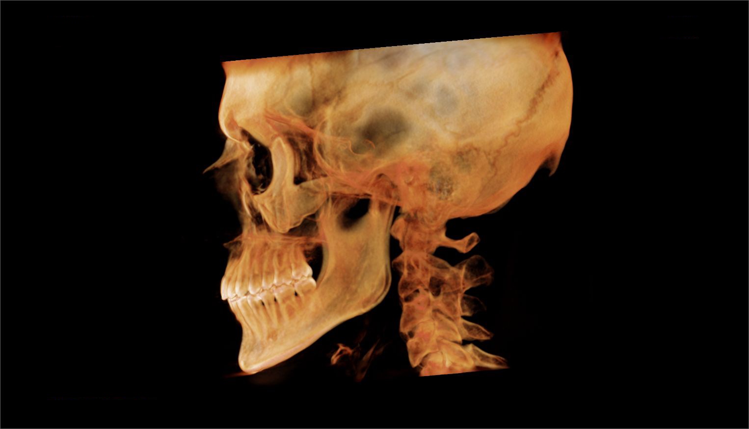
This is part of My Health, where the whole story is. This page is my airway, my tongue and my jaw.
In short: a speech-language pathologist in Massachusetts first found my tongue tie in November 2025, and in January 2026 a second one at my hospital found my tongue restricted in every direction. My myofunctional therapist says it’s one of the most restricted she’s ever seen; the ENTs have been less sure, which I write about on My Health. I’ve had two home sleep apnea tests and started a CPAP in mid-October 2026. Swallowing is difficult and painful on my right when I’m reclined with my neck extended, and a swallow study is booked for 4 November.
The April 2026 CBCT
In April 2026 I had a cone-beam CT (CBCT), a 3D X-ray of my head and neck, at a dental practice that focuses on airways. It was taken sitting up, right after the drive from Lebanon to Northwood.
The figures below annotate the practice’s exported images. Their own labels and measurements are in yellow and green; the plain-language notes were added afterwards with Claude, an AI assistant, and aren’t the practice’s read. The same scan’s view of my upper neck, figure C, is on My Spine and Brain. Tap any figure to see it full size.
Airway
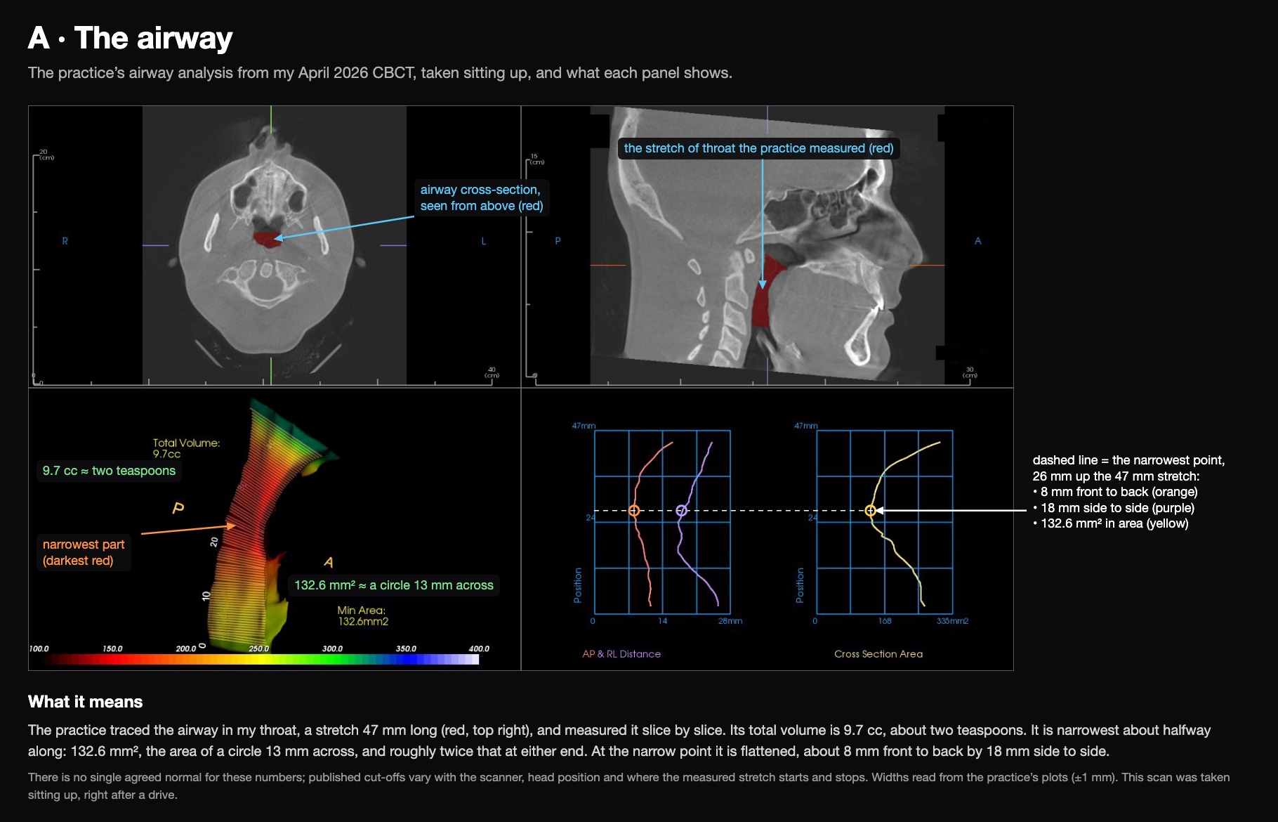
The practice traced the airway in my throat, a stretch 47 mm long, and measured it slice by slice. Its total volume is 9.7 cc, about two teaspoons. It’s narrowest about halfway along, at 132.6 mm², the area of a circle 13 mm across, and roughly twice that at either end. At the narrow point it’s flattened: about 8 mm front to back by 18 mm side to side.
Nose
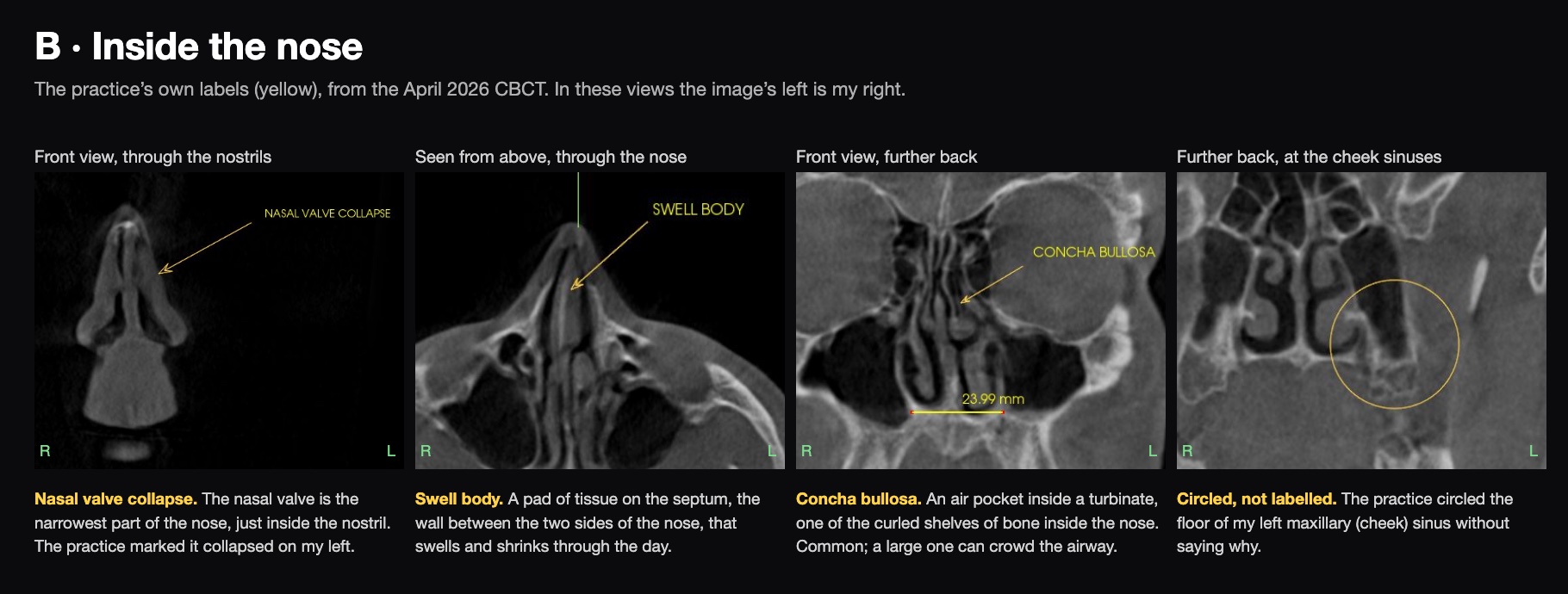
The practice marked nasal valve collapse on my left. The nasal valve is the narrowest part of the nose, just inside the nostril. They also labelled a swell body (a pad of tissue on the septum that swells and shrinks through the day) and a concha bullosa (an air pocket inside one of the turbinates, the curled shelves of bone inside the nose). They circled the floor of my left cheek sinus without a label; figure E has my best guesses at why.
Jaw
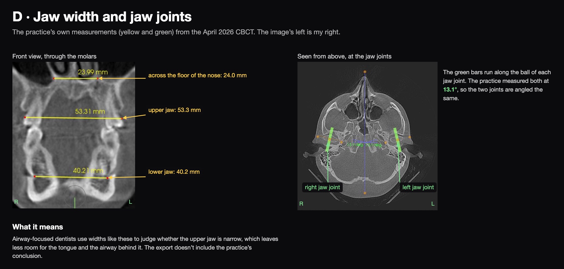
The practice measured three widths: 24.0 mm across the floor of my nose, 53.3 mm across my upper jaw and 40.2 mm across my lower jaw. Airway-focused dentists use widths like these to judge whether the upper jaw is narrow, which leaves less room for the tongue and the airway behind it. The export doesn’t include what they concluded. They measured both jaw joints at the same angle, 13.1°.
The circled spot in my left sinus
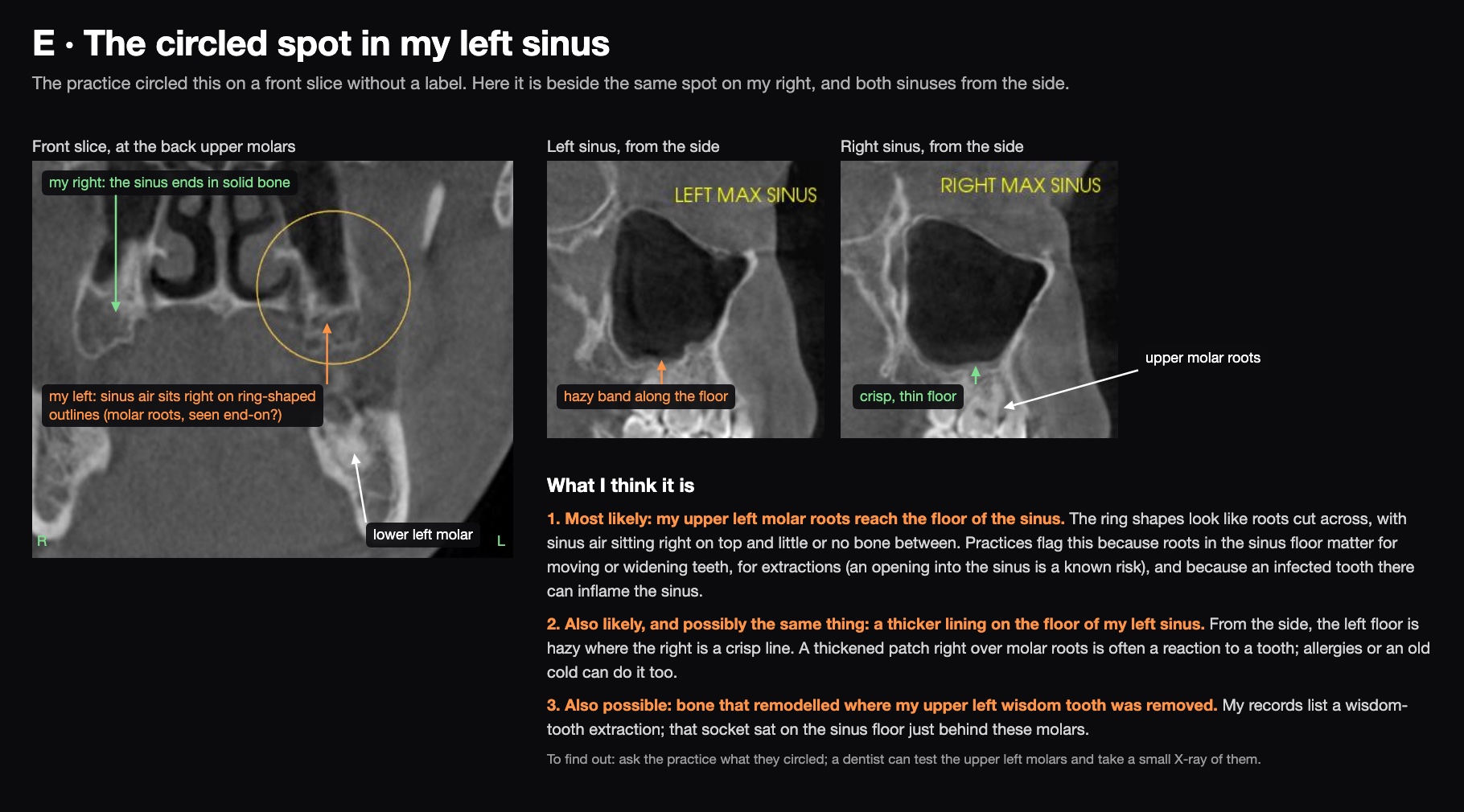
My best guess is that the roots of my upper left molars reach the floor of that sinus. In the front slice the sinus air sits right on top of ring-shaped outlines that look like roots seen end-on, with little or no bone between; on my right, the same spot is solid bone. From the side, the floor of my left sinus is also hazier than the crisp floor on my right, which looks like a slightly thickened lining over those roots. Practices flag roots in the sinus floor because they matter for moving or widening teeth, for extractions, and because an infected tooth there can inflame the sinus. It could also be bone that remodelled where my upper left wisdom tooth was removed; that socket sat on the sinus floor just behind these molars.
How my molars meet
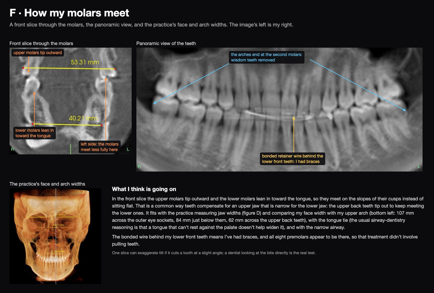
They don’t line up neatly. In the front slice through the molars, my upper molars tip outward and my lower molars lean in toward the tongue, so they meet on the slopes of their cusps instead of sitting flat, and on the left they meet less fully. That’s a common way teeth compensate for an upper jaw that’s narrow for the lower jaw: the upper back teeth tip out to keep meeting the lower ones. It fits with the practice measuring my jaw widths and comparing my face width with my upper arch (107 mm across the outer eye sockets, 84 mm just below them, 62 mm across the upper back teeth), with the tongue tie, and with the narrow airway.
The panoramic view adds two things. There’s a bonded retainer wire behind my lower front teeth, so I’ve had braces, and all eight premolars appear to be there, so that treatment didn’t involve pulling teeth.
Jaw joints in section
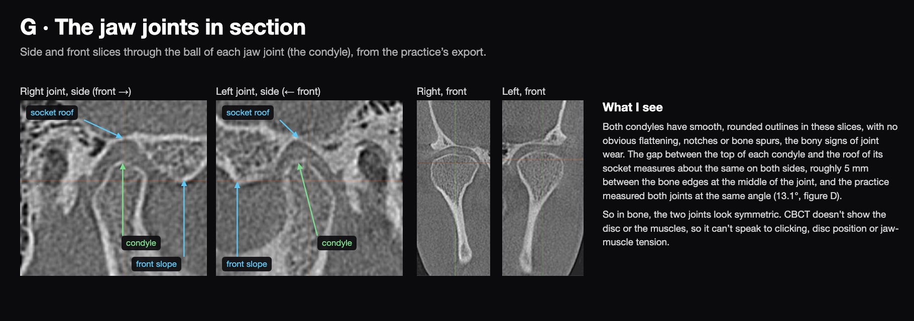
In bone, the two jaw joints look symmetric. Both condyles (the ball of each joint) have smooth, rounded outlines with no obvious flattening, notches or spurs, and the gap between each condyle and the roof of its socket measures about the same on both sides. CBCT shows bone, not the disc or the muscles, so this says nothing about clicking or jaw-muscle tension.
Side view
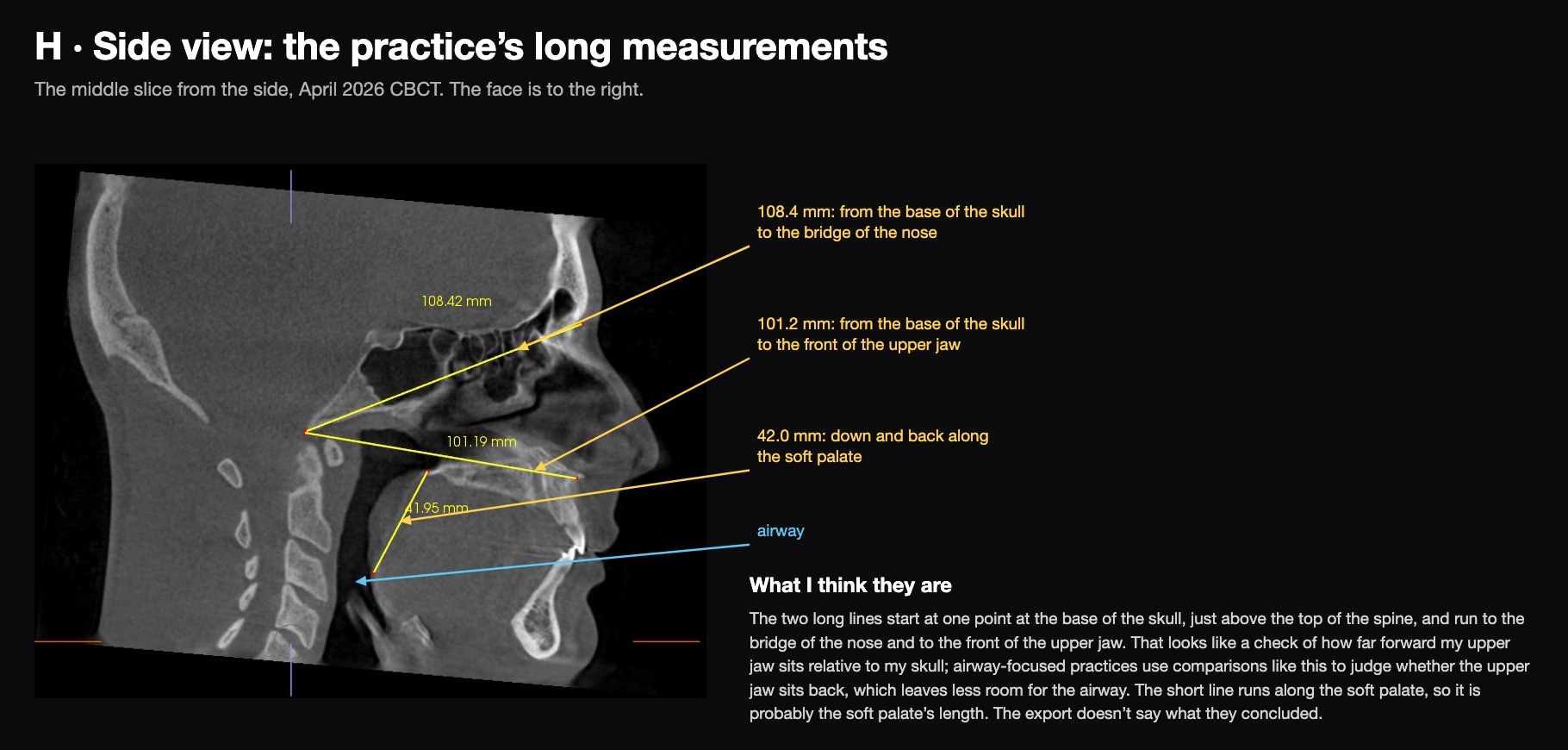
The practice drew two long lines from the base of my skull, one to the bridge of my nose (108.4 mm) and one to the front of my upper jaw (101.2 mm). That looks like a check of how far forward my upper jaw sits; an upper jaw that sits back leaves less room for the airway. The short line (42.0 mm) runs along the soft palate, so it’s probably the soft palate’s length. The export doesn’t include what they concluded.
I also have a confirmed tongue tie, and I’ve started working with a myofunctional therapist, who is concerned about both the neck rotation and possible upper airway dysfunction.