This is part of My Health, where the whole story is. It builds on My Airway, Tongue and Jaw, My Spine and Brain, How My Body Moves and the practitioners in Three Ways to Read My Case. The PRI terms are explained on How My Body Moves.
My right side feels slack and my left feels tight. This page explains fascia and the deep front line, a chain of muscle and fascia from the inner arch of the foot to the tongue, and then reads my own measurements through it: why my right half hangs, why my left pulls, and what I do about it. I worked it through with Claude, an AI assistant. Everything that applies the line to me is my reading of my own measurements, not a diagnosis and not my care team’s words.
Tap any figure to see it full size.
What fascia is
Fascia is the body’s connective tissue: sheets, sleeves and webs of collagen under the skin and around and through every muscle, organ, nerve and bone. A muscle’s outer wrapping runs on into its tendon, the tendon into the covering of the bone, and from there into the wrapping of the next muscle, so the body isn’t built from separate parts bolted together. The Fascia Research Society defines the fascial system as one three-dimensional continuum of these tissues running through the whole body.
Two things follow. Because the web is continuous, a pull in one place can pass to the next, the way tugging one corner of a bedsheet moves the far side. And fascia has nerve endings and stretch sensors in it, so it is part of how the body knows where it is.
What the deep front line is
Thomas Myers’s Anatomy Trains (2001) traces where muscles join end to end through fascia, running in one direction, into long chains he calls myofascial lines. He describes about a dozen. The deep front line is the innermost, the one he calls the body’s core. On each side it starts under the inner arch of the foot and runs up the deep calf, behind the knee and up the inner thigh, into the pelvic floor and the psoas, along the front of the spine, through the diaphragm, and up the front of the neck to the hyoid and the tongue.
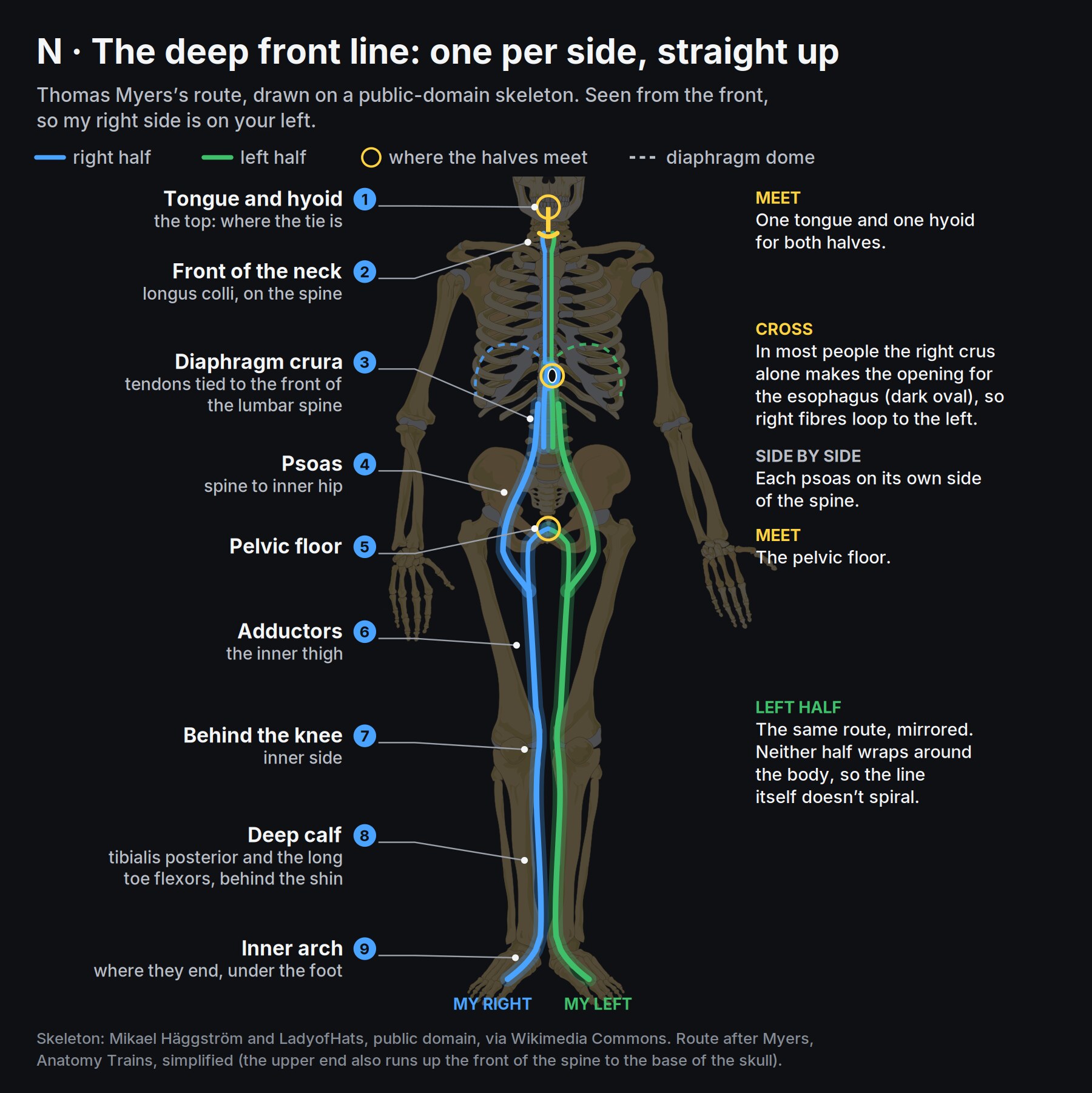
Each half runs straight up its own side. Along the inner leg it moves from back to front, from behind the knee to the front of the hip, but it never wraps around the body. The two halves meet in the middle at four places: the tongue and hyoid, the front of the spine, the diaphragm and the pelvic floor. At the diaphragm they cross: in most people the opening the esophagus passes through is made by the right half of the diaphragm alone, so right-side fibres loop around to the left.
Some of its joins are plain textbook anatomy. The diaphragm’s two legs attach to the front of the lumbar spine, and the arch it springs from beside them is a thickening of the psoas’s own fascia, so the psoas and the diaphragm are physically joined. Gray’s 1918 plates show each station:
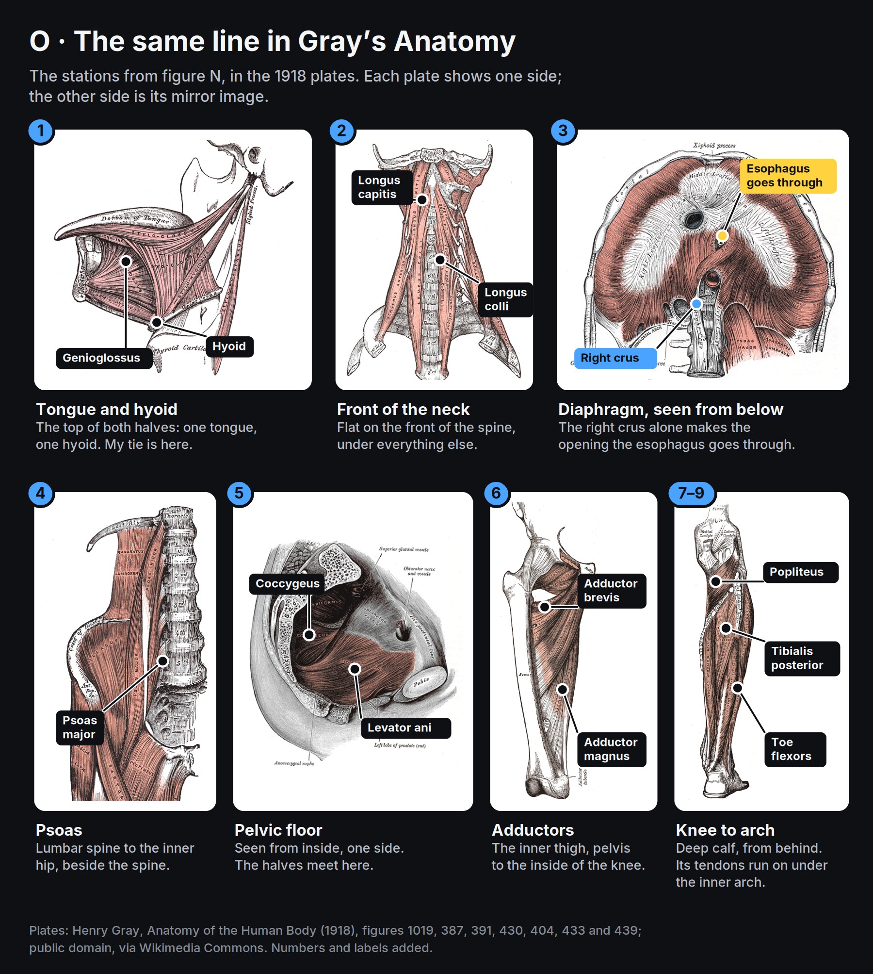
How well tested is it? In 2016, Jan Wilke and colleagues reviewed the cadaver dissection studies for six of Myers’s lines. Three were well supported, two in part, and one, the superficial front line, not at all. The deep front line wasn’t among the six: it rests on Myers’s own dissections and on joins like the psoas and the diaphragm. Dissection also shows only that tissues connect, not how much force passes along them in a living body. So I use the line as a map for reasoning about my measurements, not as a proven mechanism.
Where my twist fits
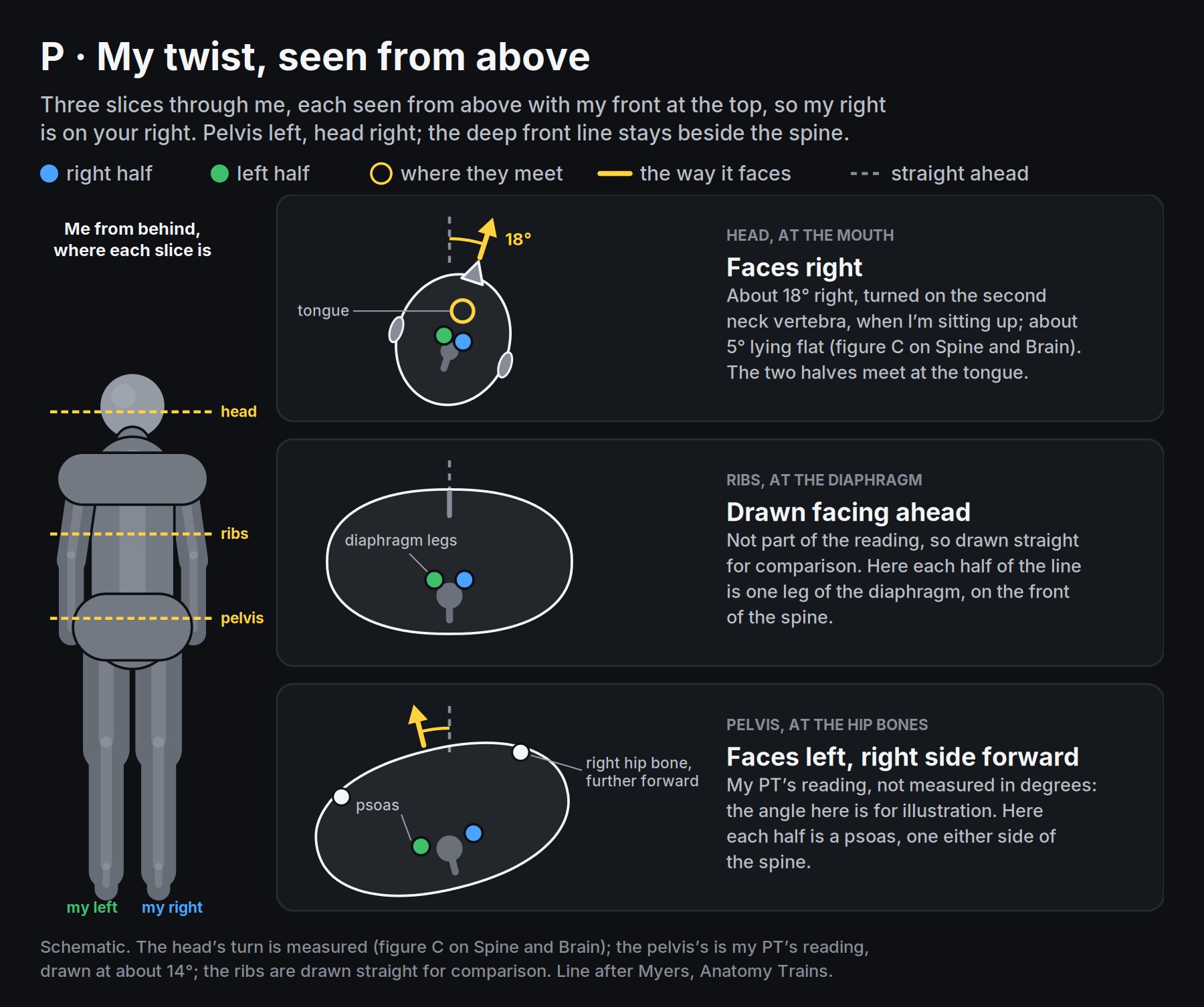
The twist I feel is real and measured, but it belongs to the whole body, not to this line. My pelvis faces left with its right side forward, and my head is turned about 18° right at the second neck vertebra when I’m sitting up (figure C), against about 5° lying flat. Hips one way, head the other, like being stopped mid-stride. The lines that wrap around the body are others: Myers’s spiral line, from the skull across the upper back to the opposite shoulder, across the belly to the hip and under the arch, and his functional lines, from each shoulder to the opposite hip. The deep front line sits inside that twist.
What’s shared and what’s only on the right
Two limits sit on the midline, so they affect both halves, and both are older than the pattern. The right-only ones are newer or local.
| Limit | Side | Since |
|---|---|---|
| Tongue tie | Midline: both halves | All my life |
| Weak pelvic floor squeeze | Midline: both halves | A 2024 pelvic floor test, before the injury (my records) |
| Palate narrower on the right, and my tongue doesn’t reach that side | Right | My myofunctional therapist’s palate scan, October 2026 |
| Less feeling in my heel, weaker ankle and toes | Right | The Romanian deadlift injury, April 2025 |
| The pattern itself | None of it existed before the injury |
Why the right hangs and the left pulls
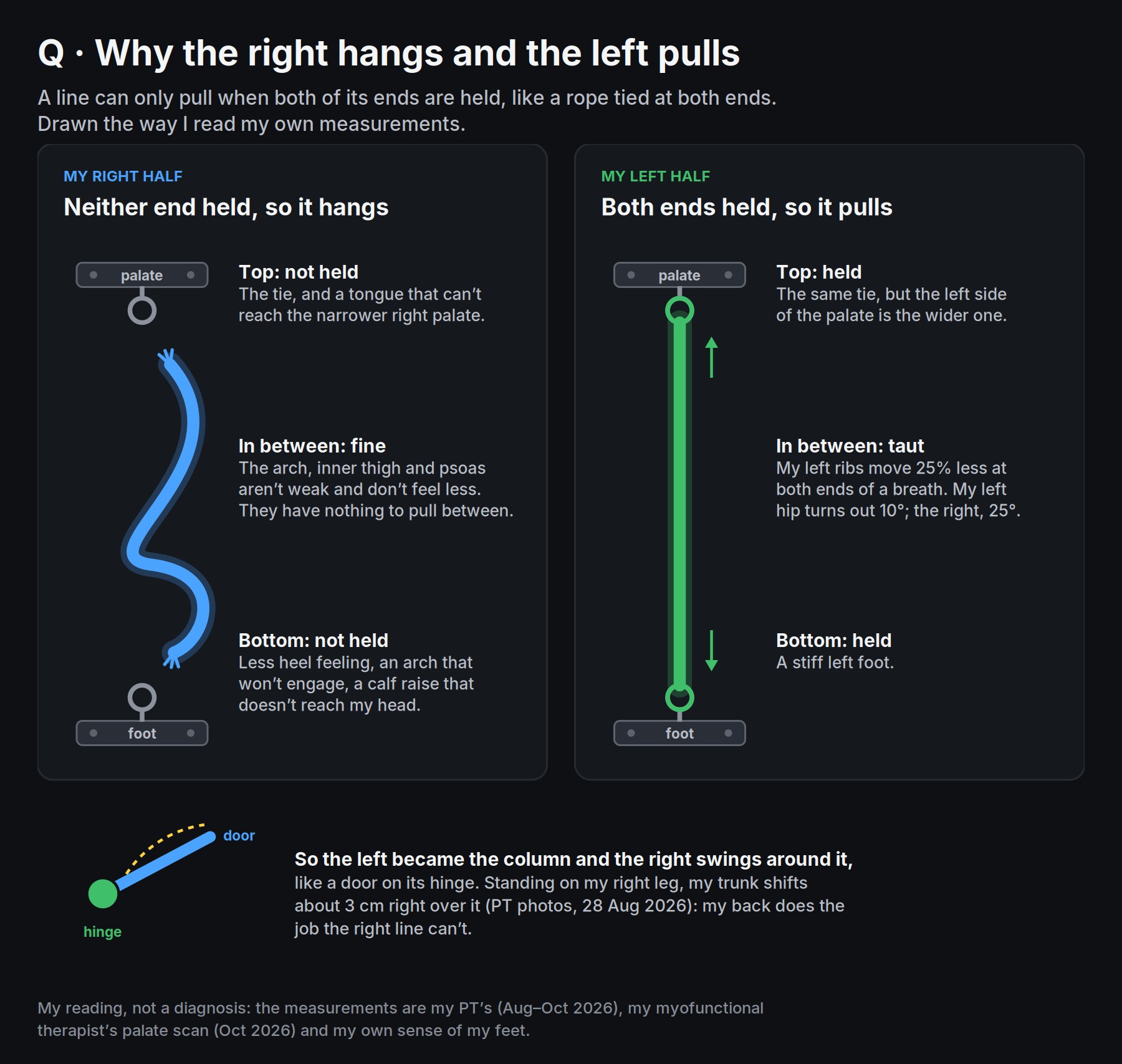
A line can only carry force when both of its ends are held. A rope tied at one end hangs; tied at both, it can pull.
My right half has neither end. The bottom is unreliable: less feeling in my heel, an arch that won’t engage, a calf raise whose push doesn’t reach my head. The top is partly missing: the tie, and a tongue that can’t seat on the narrower right side of my palate. With nothing to pull between, it hangs. That fits how it feels: force doesn’t carry up my right side, yet the right isn’t weaker and doesn’t feel less. The muscles are fine; the anchors aren’t.
My left half has both. My left foot is stiff, my left ribs move 25% less at both ends of a breath, and my left hip turns out 10° against the right’s 25°. The body has to stand on something, so the left became the column and the right swings around it, like a door on its hinge. Standing on my right leg, my trunk shifts about 3 cm right over it: my back does the job the right line can’t.
The neck is the top of the twist, not its source. Turning the head right is the job of the left sternocleidomastoid and the left upper trapezius, and both are my more developed side. The turn is about 18° sitting up and about 5° lying flat, so it is mostly a response to being upright, not fixed bone. That’s why I leave my neck alone: it should follow the base.
Which end started it
The timing points at the bottom. The tie and the weak pelvic floor were there before, without this pattern. The right-sided pattern appeared after the April 2025 injury hit the bottom of the right line. My best reading: the tie set a low ceiling for both halves, the injury took the right’s bottom anchor, and my body reorganised around the left. So both ends need work.
One caveat. My EMG was normal (9 October 2026), the S1 nerve roots look free on my own read of my October lumbar MRI (the report is still pending), and I can do a pistol squat on either leg. The heel, the calf and the big-toe flexor are supplied mainly by S1 and S2, but the main arch muscle, tibialis posterior, is mostly L4 and L5. So “the injury took the anchor” explains the heel and the calf, not the whole arch, and it is about sensing and timing, not strength.
What I do about it
Everything here comes from the rope in figure Q. My right half can’t pull because nothing holds its ends, and making the rope stronger or longer won’t change that. So the work is to give it ends: first ends the outside world holds for me, then ends I hold myself.
A held end is something that stays put when the line pulls on it. At the bottom, it’s whatever my right foot or right hip is pressed against. At the top, it’s my tongue sealed to the roof of my mouth. With both held, a push or a breath has a path from one end to the other. With neither, my left side does the job instead, and that is the pattern I’m trying to undo.
So for any position or exercise, I ask one question: what is holding the right half’s ends here? If the honest answer is “nothing”, it’s too early for that one, however easy it looks. I add support until something is.
That question sets the order. Each stage takes away a bit of outside support, so the right half has to do more of the holding itself:
| Stage | What holds the bottom end | What the right half has to do |
|---|---|---|
| Now: lying on my right side | The floor, along the whole right side | Only breathe against it, so it can feel where it is |
| Next: the same, right foot to a wall | The wall, under my foot | Push from one fixed point and let the push travel up |
| Later: part-way up, on my right forearm | The floor, under my right hip and forearm only | Hold my trunk up between two small contacts |
| On hold: standing on my right foot | My own foot: the heel and the balls of the big and little toes | Make the anchor itself, then pull from it |
Standing is on hold because I can see it isn’t ready: on my right leg my trunk shifts about 3 cm right, which is my back doing the holding, not my foot.
The question works outside the exercises too. At a desk with my right foot flat and some weight in the heel, the floor holds the bottom end; with my feet crossed or tucked under the chair, nothing does.
Four more rules sit alongside:
- The breath is the line’s own movement. On a relaxed breath out, the diaphragm domes up and the pelvic floor and tongue rise with it. The work is breathing in positions, not stretching the line.
- Don’t stretch or release the left. It’s the only column holding me up. It will let go when the right can take load, not before.
- Don’t strengthen the right’s stations, the arch, inner thigh and psoas. They aren’t weak; they have nothing to pull from.
- One change at a time, kept only while its check moves.
The stages follow Bill Hartman’s phases: lying on the side, then part-way up, then upright. The push into a wall is from Reconsider… Rolling Patterns with Bill Hartman, 19:12, where he uses it to drive inward rotation through the right side.
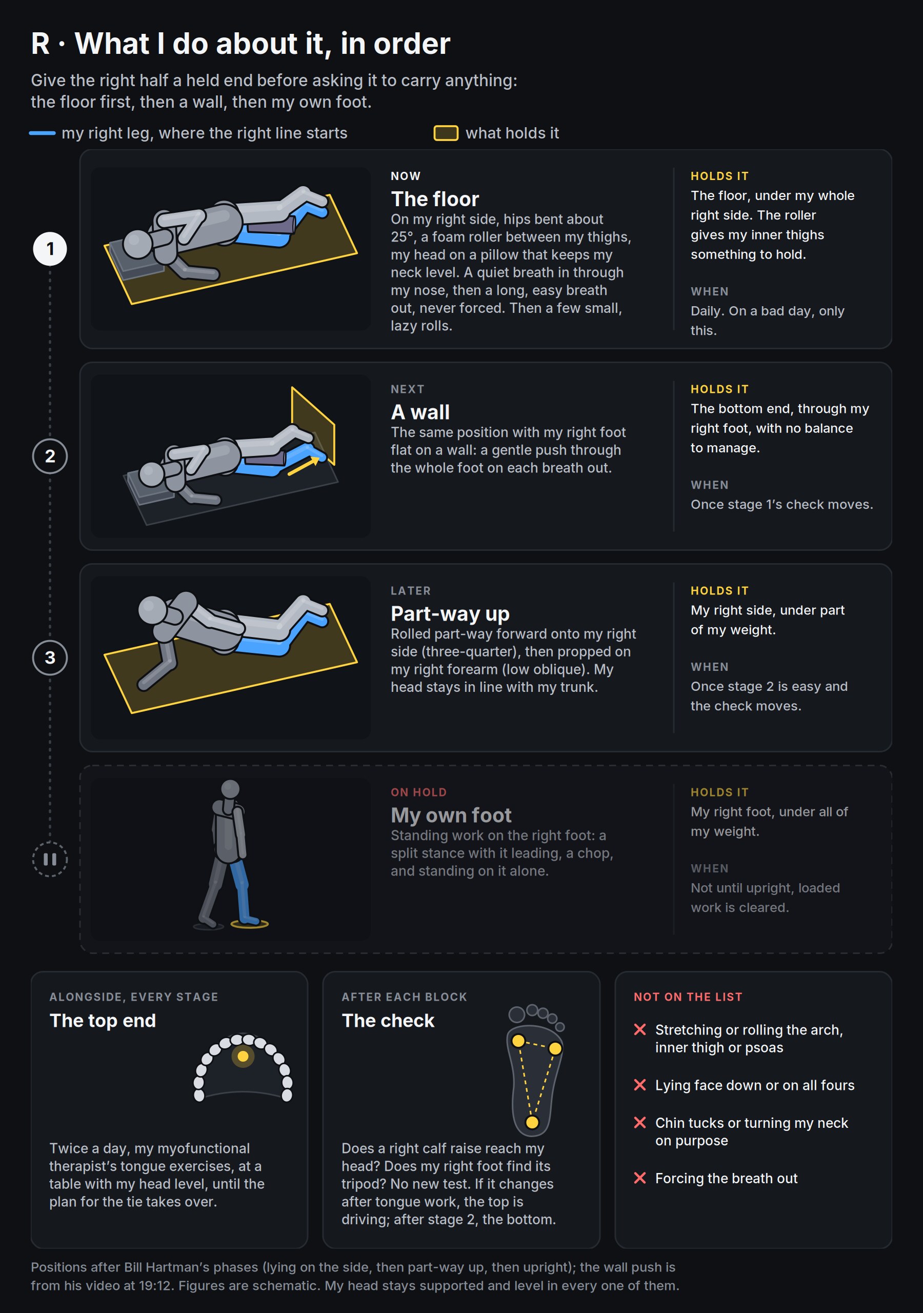
The top end follows the same logic. Alongside every stage, I do my myofunctional therapist’s tongue exercises twice a day at a table, with my head level, until the plan for the tie takes over: they teach my tongue to hold the palate by itself, so the line has something to pull toward. Stage 1 is daily, and on a bad day it’s the only thing I do.
After each block, one check, and no new test: does a right calf raise reach my head, and can my right foot find that three-point stance? If it changes after tongue work, the top is driving; if it changes after stage 2, the bottom is. I note anything else that day, like sleep or a PT visit, before crediting either.
Not on the list: stretching or rolling the arch, inner thigh or psoas; lying face down or on all fours; chin tucks or turning my neck on purpose; forcing the breath out.
Other people’s cases
On 1 November 2026 I searched for published cases like mine. None matches the whole picture, a tongue tie with a rotated upper neck, a right TMCC and a left AIC, and none of the near matches reports a full recovery.
| Case | How it matches | What happened |
|---|---|---|
| r/Posture, 29F, October 2026 | Left AIC, both BCs, right TMCC; a tongue that drops; poor sense of the torso; “fight or flight” | 14 years of chiropractic, PT, PRI, orthotics and release work. A right arch insert helped stability, and fixing a left rib flare stopped the pain spreading. The right TMCC and core pain persist. No tie found |
| r/Posture, January 2023 | About a year of PRI, then a tie release | The TMCC went neutral and the neck “freed up”; the right BC, and no air into the right side of the back, persisted |
| r/Posture, 21M, July 2022 | Right BC and left AIC, a tie, a narrow palate and airway, jaw pain | Planned a splint, expansion and a release. No outcome posted |
| “Alex”, an orthodontic journal case, 2020 | Forward head, mouth breathing, a tie | A release plus PT improved his posture, then it relapsed when the frenum reattached, with scarring and too little tongue retraining. A second release, daily tongue work and palate expansion over 26 months: head upright, breathing through the nose. No measurements |
| A 14-year-old, the same journal, 2023 | Less right neck rotation, forward head, a tie | Chiropractic and craniosacral work around a release; better sleep, fewer headaches. No measurements |
| Zaghi and colleagues, 2019, 348 people surveyed | A tie, no posture data | Release plus myofunctional therapy: 91% satisfied, 78% less mouth breathing, 78% less muscle tension. A look-back survey, not a trial |
| Boyle, 2011, 65F | Left AIC, low back and sacroiliac pain, no top end | Eight weeks of left-AIC repositioning and stabilisation: disability score from 20% to 0, left hip inward rotation from 40° to 45° |
What they add: where a tie was involved, improvement came from the release first and retraining after, and the one documented relapse came from neglecting the retraining. The two cases that treated the neck directly have no measurements and can’t separate the neck work from the release, so they don’t change my “leave the neck alone”. Boyle’s exercises reposition the left AIC rather than stretch the left or load the right’s stations, so they fit the principles above. The right arch insert in the first case looks like the bottom anchor found by accident, but it is one anecdote.
Sources and image credits
- Fascia and the fascial system, as defined by the Fascia Research Society’s nomenclature committee: Fascia Research Society.
- The deep front line: Thomas Myers, Anatomy Trains.
- How well Myers’s lines hold up in dissection, and fascia’s nerve endings: Wilke, Krause, Vogt and Banzer, “What Is Evidence-Based About Myofascial Chains: A Systematic Review”, Archives of Physical Medicine and Rehabilitation 97:454–461 (2016), doi:10.1016/j.apmr.2015.07.023.
- The esophageal opening made by the right half of the diaphragm, in cadaver and CT studies: Folia Morphologica, Advances in Clinical and Experimental Medicine.
- The big-toe flexor’s nerve roots, mainly S1 and S2: Radiopaedia, Kenhub.
- The cases: linked in the table. Zaghi and colleagues, Laryngoscope Investigative Otolaryngology 4(5):489–496; Boyle, Physiotherapy Canada 63(2):154–163.
- Figure N’s skeleton: Mikael Häggström and LadyofHats, public domain, via Wikimedia Commons.
- Figure O’s plates: Henry Gray, Anatomy of the Human Body (1918), figures 1019, 387, 391, 430, 404, 433 and 439, public domain, via Wikimedia Commons.
- The figures were drawn with Claude. They are schematic, and the route is simplified from Myers.


