This is part of My Health, where the whole story is. This page is how my body moves and holds itself: what my physical therapist found, the range-of-motion “chessboard”, what my standing photos measure, and my eyes, which shape how I hold my head.
What my physical therapy found
From May to late September 2026 I worked with a physical therapist who is certified by the Postural Restoration Institute (PRI) but doesn’t work from PRI’s model. Their mentor is Bill Hartman, whose model they trust, which is why I’ve studied Hartman’s teaching (Three Ways to Read My Case).
In PRI’s terms I’d been describing myself as a Left AIC with a Right BC, overlaid with a Right TMCC, and I built my daily routine and training around that. The terms are explained below.
- Ribs: in August 2026, my PT assessed my ribs as compressed rather than flared.
- Reflexes: primitive reflex testing that month showed my elbows bending when I turn my head side to side on all fours.
My therapist doesn’t read me through PRI, and of the three patterns, only two fit my measurements. The Right TMCC and the Right BC do: my head sits turned right on C2, the torsion Hallinan and Mallon say locks the neck into a Right TMCC (Three Ways to Read My Case), and my right shoulder sits lower in all 12 standing photos, turns inward least of all (5°), and feels like it’s going to fall off. The Left AIC fits less well. My left thigh does sit turned in, but as I read my standing photos below, my right pelvis sits forward, the opposite of a textbook Left AIC, and both hips turn inward only 5°, which is locked on both sides and closer to what PRI calls a Patho PEC. PRI’s tools can still be useful to me even where its labels don’t fit.
What the PRI terms mean
PRI, the Postural Restoration Institute’s approach, starts from the body being lopsided by design: the liver sits on the right, the heart on the left, and the right side of the diaphragm is bigger and stronger. Most people lean into that and stand more on their right leg. PRI names the chains of muscles that hold a person in that lean, and calls a chain stuck when it can’t let go.
- AIC, the anterior interior chain: the deep chain from the diaphragm down through the hip flexors to the outside of the thigh. A Left AIC tips the left side of the pelvis forward and turns the pelvis toward the right, with the weight on the right leg. PRI treats it as the pattern nearly everyone has to some degree.
- AF IR: the thigh turned inward in the hip socket. A left thigh that lives turned in (left AF IR) is the hallmark of a Left AIC.
- BC, the brachial chain: the chain from the diaphragm and the front of the rib cage to the shoulder blade and arm. A Right BC holds the right rib cage and shoulder down and forward.
- TMCC, the temporal mandibular cervical chain: the jaw and neck muscles on one side. A Right TMCC is that chain stuck on the right, which changes how the head sits on the neck and how the teeth meet.
- PEC, the posterior exterior chain: the back muscles on both sides. When the one-sided pattern gets locked in, both sides of the pelvis tip forward and neither hip can turn inward. PRI calls that a Patho PEC: stuck on both sides instead of lopsided.
PRI’s own material is at posturalrestoration.com, written for clinicians.
The chessboard
My physical therapist tracked my ranges of motion on a “chessboard”, a set of measurements from Bill Hartman’s model. This is the retest from 24 September 2026, after a breathing and positioning intervention, in the hardest week of my year.
| Measure | Left | Right |
|---|---|---|
| Straight leg raise | 40° | 45° |
| Pelvic reorganization / hip flexion | 75° | 75° |
| Hip external rotation | 10° | 25° |
| Hip internal rotation | 5° | 5° |
| Late hip internal rotation | −20° | −25° |
| Late hip external rotation | 35° | 30° |
| Shoulder flexion | 70° | 75° |
| Shoulder external rotation | 85° | 85° |
| Shoulder internal rotation | 10° | 5° |
| Late shoulder external rotation | −5° | 0° |
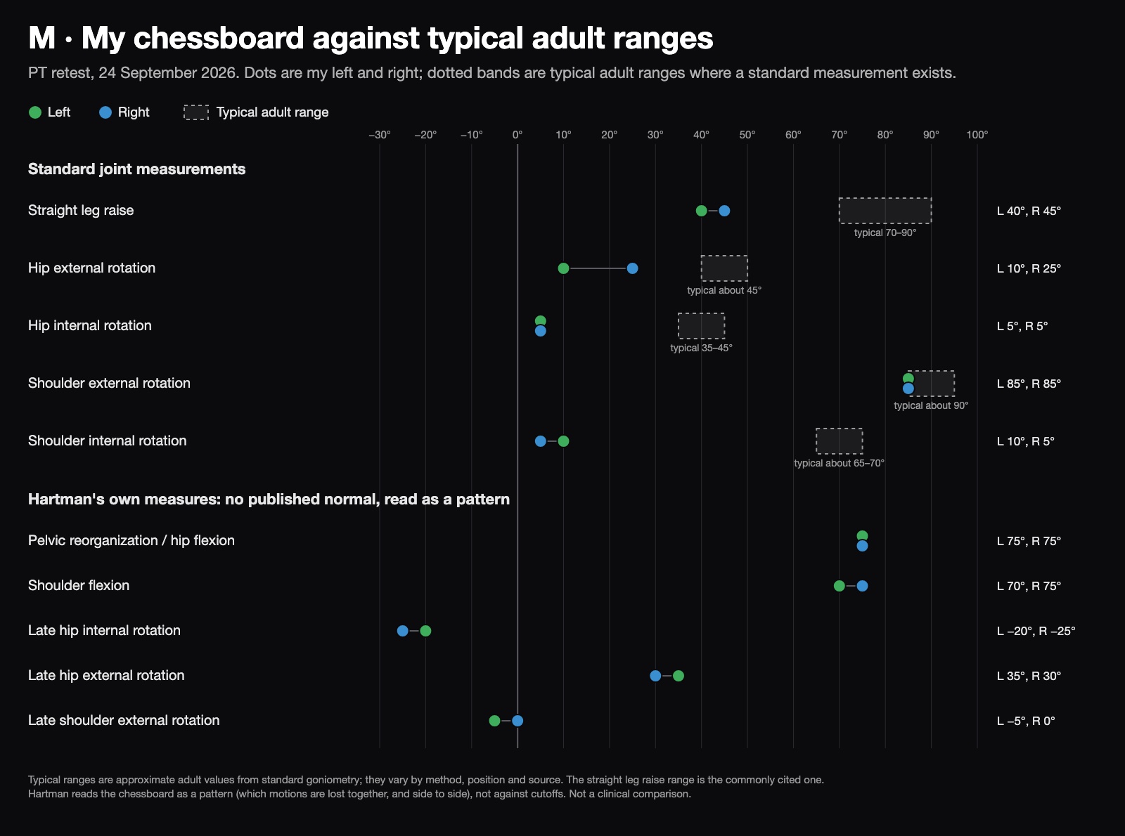
Where a test matches a standard joint measurement, the dotted band shows the typical adult range. My hips and shoulders turn inward only 5–10°, against a typical 35–45° at the hip and about 65–70° at the shoulder. My straight leg raise is 40–45°, against a typical 70–90°. Shoulder external rotation is the one near typical. Hartman’s own measures have no published normal, so they’re plotted without one.
What I read in it:
- Almost everything is restricted, internal rotation most of all: 5° at both hips, 5–10° at the shoulders, and a straight leg raise of only 40–45°. That’s close to word for word what Hartman calls narrow-ISA “end game” compression: both hip internal and external rotation lost, the glutes and adductors clamping from outside, and a poor straight leg raise (End Game, 1:00).
- My left hip is the most stuck, turning out 10° against 25° on the right. My left thigh lives turned inward, the same thing my standing photos show as the left knee drifting in.
- My right shoulder has the least internal rotation, 5°. It’s my throwing shoulder.
- A neck input changed the picture. This retest came after a 4-inch half-moon bolster under my neck produced substantial changes in these ranges. I don’t have the numbers from before it.
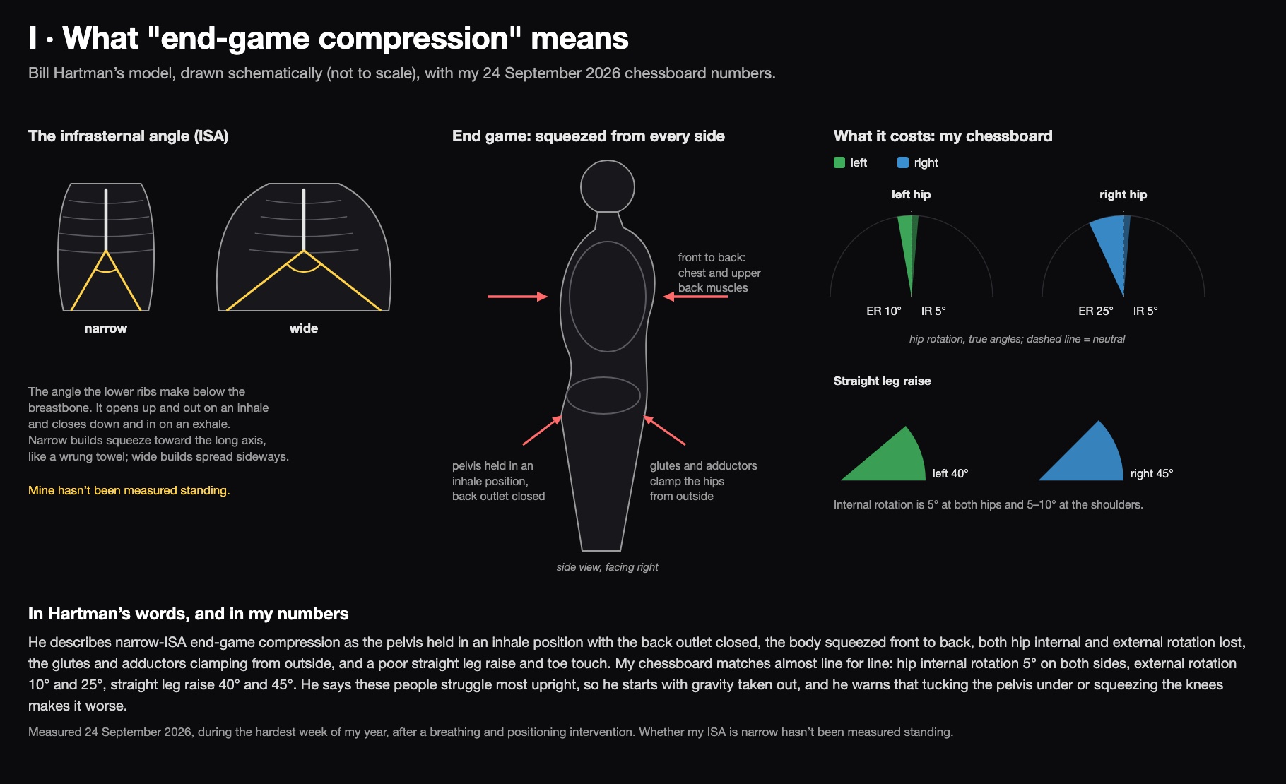
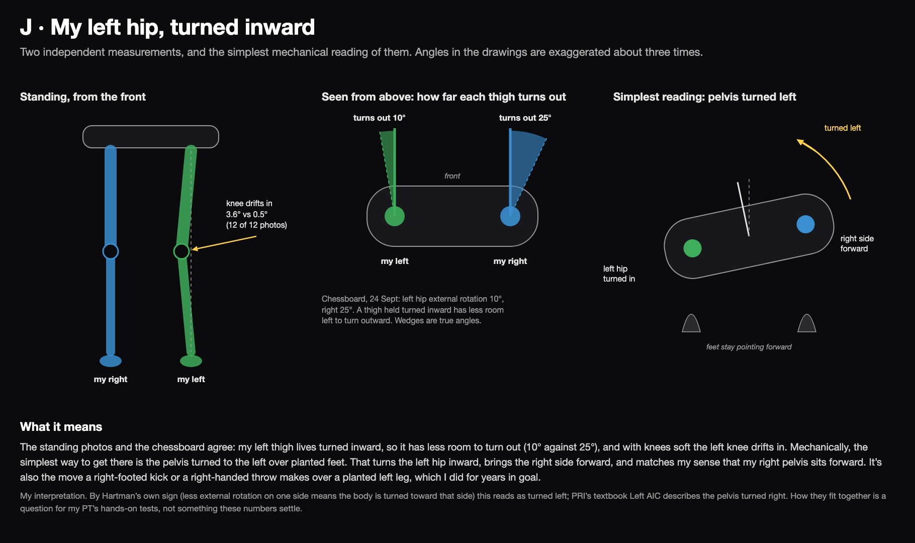
My reading of the hip: the simplest way to get a left thigh that lives turned inward is a pelvis turned to the left over planted feet. That also brings my right side forward, which is exactly where I feel my right pelvis sits. PRI’s textbook Left AIC has the pelvis turned the other way, so this is a question for my PT’s hands-on tests.
What my standing photos measure
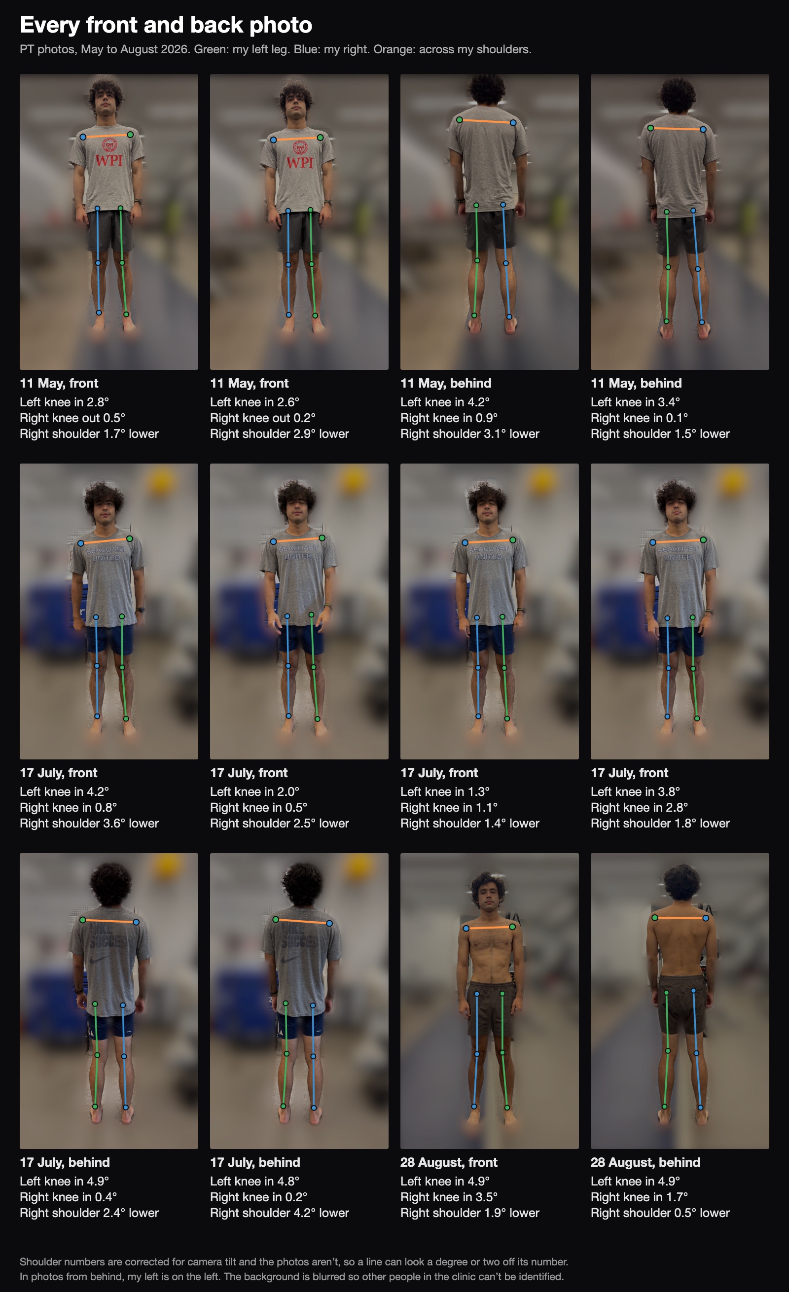
I measured 12 front and back photos from my PT sessions between May and August 2026, using a small tool I built on Apple’s on-device body-pose detection. It corrects for the camera being tilted. To separate my body from the camera, I used a simple test: a real asymmetry points to the same side of me whether the photo is taken from the front or the back, while a camera artefact points to the same side of the image. Two findings passed:
- My right shoulder sits about 2° lower (2.3° on average), in all 12 photos.
- My left knee drifts inward about 3.7°, against about 1° on the right, and further than the right in all 12.
Here is every one of those photos, with what the tool measured in each:
From the side, my pelvis sits a few centimetres ahead of my ankles and my upper body leans back about 5–7°. I stand by hanging on my hips rather than holding myself up. Everything else either stayed within the noise or turned out to be the camera: pelvis level, leg length and sideways shift. These numbers are small. Plenty of people without symptoms show this much, and neither changed between May and August.
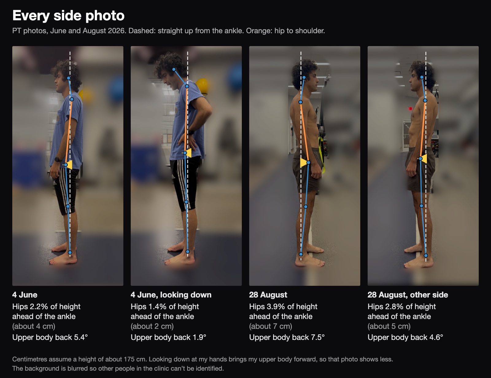
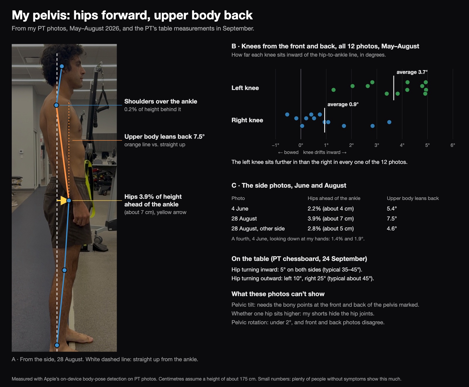
The white dashed line runs straight up from my ankle. My shoulders sit right on it and my hips sit ahead of it, and the orange line from hip to shoulder shows my upper body leaning back to balance. In the dot plot, each dot is one photo; the further right, the further the knee sits inward. What the photos can’t show is how my pelvis is tilted, so they can’t say whether my lower back curve is bigger than normal. That takes the bony points at the front and back of the pelvis marked on a side photo.
My reading: I’m right-handed, and I played in goal, throwing overhand and taking goal kicks with my right leg. A dominant throwing shoulder commonly sits lower and feels looser, and my right shoulder does feel lax. Every right-footed kick or right-handed throw also turns the pelvis to the left over a planted left leg, which rotates the left hip inward. That’s the position PRI calls left AF IR, the hallmark of Left AIC, and a left knee that drifts in is what it looks like standing. So years of right-sided sport fit both numbers.
My eyes
I’ve known about my XLRS, a genetic retinal condition, since I was eight or nine, and it’s been tracked ever since. Retinal testing at Mass Eye and Ear in March 2026 confirmed it in both eyes, worse on the right: my right eye’s field of view is narrowed, with blind spots, while my left eye’s field is normal. I’ve relied on my left eye for as long as I can remember, and I’m very nearsighted.
Here’s what that looks like in three dimensions, as a sphere around my head, one eye at a time. My left eye’s field is normal. My right eye’s is smaller, with three blind spots: one near the centre, one up and toward my nose, and one outward to the right. Only my right eye can see the far right, which is where it loses the most. That’s a sketch, not a measurement: the report says where the field is lost but not by how many degrees, so I’m asking Mass Eye and Ear for the field maps.
Left eye
Right eye